J Comput Assist Tomogr 1994 Sep;18(5):737-744
Chronic eosinophilic pneumonia: evolution of chest radiograms
and CT features.
Ebara H, Ikezoe J, Johkoh T, Kohno N, Takeuchi N, Kozuka T, Ishida
O
Department of Radiology, Osaka University Medical School, Japan.
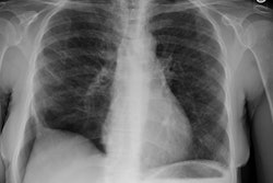
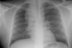
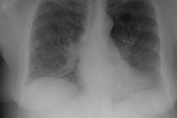
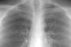
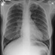
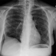
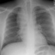
OBJECTIVE: Our object is to describe and compare the findings on plain chest radiographs and CT scans in patients with chronic eosinophilic pneumonia of varying duration, as judged by their clinical history. MATERIALS AND METHODS: We retrospectively reviewed the initial chest radiographs and initial CT scans that were obtained before treatment with corticosteroid in 17 patients with pathologically proven or clinically diagnosed chronic eosinophilic pneumonia. RESULTS: Eleven of the 17 patients showed predominantly peripheral patchy or confluent consolidation with or without ground-glass opacities on chest radiography. Sixteen patients, on the other hand, showed various types of abnormalities with peripheral predominance on CT. The seven patients in whom the initial CT was performed within 1 month after the onset of symptoms had dense confluent consolidation (7/7) with or without ground-glass opacities. When the initial CT was performed 1-2 months after onset of symptoms, inhomogeneous patchy consolidation or nodules (5/7) or ground-glass opacities (2/7) were observed. When the initial CT was performed > 2 months after the onset of symptoms, streaky or band-like opacities (1/3) or lobar atelectasis (1/3) was seen. CONCLUSION: Patients with chronic eosinophilic pneumonia show an evolution of CT features at varying time intervals after the onset of disease.
PMID: 8089322, MUID: 94375686
