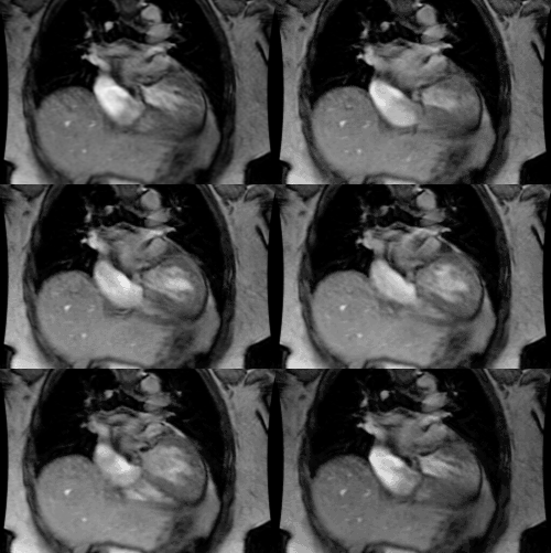
Williams Syndrome (Williams-Beuren syndrome):
Clinical:
Williams syndrome is a rare genetic dysmorphic syndrome
characterized by supra-aortic
valvular stenosis, peripheral pulmonary stenosis, and
life-threatening coronary
artery obstructive lesions (in 5-10% of cases). These defects
occur in
association with an elfin-like facies, mental
retardation/developmental delay, behavioral changes, and neonatal
hypercalcemia [2]. The disorder is caused by a microdeletion at
7q11 on chromosome 7 [1,2]. The affected region of this chromosome
includes the elastin gene, which encodes for elastin protein [2].
The ensuing decrease in the synthesis of elastin leads to a number
of histologic changes in the vascular wall including decreased
elasticity, thickening of the tunica media, and smooth muscle cell
hypertrophy [2]. These changes lead to the characteristic
supravalvular aortic stenosis, hypoplasia of the aortic arch, and
pulmonary artery stenosis [2].
Supravalvular aortic stenosis is the most common cardiovascular
manifestation f the disorder (70% of patients) and is rarely seen
outside of this patient population [2]. The lesion is most
commonly located at the sinotubular junction superior to the
aortic valve producing an "hourglass" appearance to the ascending
aorta [2]. If moderate-to-severe stenosis is left untreated, it
may result in significant LV hypertrophy or heart failure during
the first 5 years of life and surgical intervention is often
warranted [2].
Supravalvular aortic stenosis is associated with an increased
risk of sudden cardiac death, likely the result of alterations in
coronary perfusion [2]. In some patients, the aortic valve
leaflets fuse with the supravalvular ridge ("coronary hooding"
limiting blood flow into the coronary arteries and causing
myocardial ischemia [2]. Also- because the coronary ostia are
located proximal to the region of stenosis, they are subjected to
abnormally high systolic pressures [2]. The increased hemodynamic
stress may lead to fibrosis and smooth muscle hypertrophy withi
the vessel wall, resulting in coronary stenosis [2]. Sudden death
can occur under general anesthesia in these patients- most likely
as a result of a drop in coronary perfusion pressure [2].
Therefore, consideration should be given to imaging these patients
without sedation [2].
Hypoplasia/stenosis of the aortic arch (more commonly) or
descending thoracic aorta can also occur [2]. Mesenteric or renal
artery stenoses can also be seen [2].
Pulmonary artery stenosis can be found in up to 45% of patients
[2]. The stenosis can be discrete or long segment [2]. The
pulmonary artery stenosis often regresses or resolves with time
and patients with PAS can be managed conservatively [2]. If
treatment is required, balloon angioplasty can be performed for
short segment stenoses, but surgery is often necessary for
long-segment disease or discrete stenosis in the main pulmonary
artery [2].
REFERENCES:
(1) J Nucl Cardiol 2003; Grossfeld PD. The genetics of congenital
heart
disease. 10: 71-76
(2) J Cardiovasc Comput Tomogr 2013; Gray JC, et al. Cardiovascular manifestations of Williams syndrome: imaging findings. 7: 400-407


