Taking care of patients in the midst of a pandemic can require a little ingenuity as well as science. During the COVID-19 outbreak, hospitals have developed creative solutions to provide x-ray services in a way that keeps both patients and personnel safe.
These solutions can range from the low-tech -- such as glass barriers to separate patients from radiology personnel -- to the high-tech -- using artificial intelligence (AI) algorithms to analyze medical images.
With either approach, imaging professionals are sharing their experiences and research findings with the radiology community to maximize their ability to battle the COVID-19 pandemic.
X-ray behind glass
In an effort to minimize the risk of infection, a number of radiology departments have reorganized their operations to reduce contact between staff and possible carriers of the SARS-CoV-2 virus. Patient intake areas in some cases have been located in temporary facilities outside the radiology department. And within departments, patient and staff routing has been changed to minimize interaction.
One increasingly common solution takes advantage of the fact that x-ray beams are not significantly attenuated by glass. At the same time, mobile x-ray systems offer flexibility in where images can be acquired -- a flexibility not available with big iron modalities like CT and MRI.
A group at Alfred Health in Melbourne, Australia, got creative in developing an approach that allows the x-ray unit to remain outside an isolated patient's room -- conserving personal protective equipment (PPE), reducing radiographer risk, and speeding the diagnostic process. They published details about their technique on July 13 in Physical and Engineering Sciences in Medicine.
With the technique, the radiographer inside the patient area positions the bed close to the glass door and places the digital detector behind the patient for an erect anterior-posterior chest x-ray before stepping away from the patient during x-ray exposure. The radiographer outside the area positions the x-ray tube head close to the glass door and steps laterally away to initiate the x-ray while maintaining visual contact with the patient, according to Zoe Brady, PhD, chief physicist in the hospital's radiology and nuclear medicine department.
"Depending on a patient's room configuration, the through-glass technique can follow a patient through their (hospital) journey," Brady wrote in an email to AuntMinnie.com.
The technique can be used as part of an initial assessment in the emergency department. In the intensive care unit, chest x-rays can monitor changes or the insertion of new lines, such as an endotracheal tube if the patient needs intubation, a nasogastric tube for feeding, or a central line for drug administration. Then, to monitor for ongoing complications, x-rays can be acquired in the wards as the patient recovers, according to Brady.
"Taking the x-ray unit to the patient reduces the number of patient contacts and limits movement through the hospital, not just [in] the radiology department but [in] lifts, corridors, [and on] door handles touched by the orderly," Brady said.
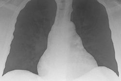
Mobile x-ray systems have huge advantages, especially for infection control, noted Dr. Danielle Toussie of Mount Sinai Hospital in New York City. Toussie has led research showing that chest x-rays acquired when patients present in the emergency room with COVID-19 symptoms can predict the severity of the disease.
"When there are large numbers of patients with COVID positivity in the emergency department, restricting these patients from being transported throughout the hospital is hugely important," Toussie noted.
Additionally, bringing portable x-ray to the patient means not having to disinfect an x-ray room, saving time, resources, and money, she explained in an email to AuntMinnie.com.
Another facility has also leveraged mobile x-ray to good use in managing patients with COVID-19. Staff and administrators at Penn State Health Radiology at Milton S. Hershey Medical Center recognized that the time-consuming nature of imaging COVID-19 patients could hinder their ability to image patients with other conditions.
Their solution was to convert a storage area into a radiology annex, with an external door that's separate from the one into the main radiology department. A mobile x-ray system is positioned in the doorway, with patients approaching the system from outside the building.
A piece of plexiglass was bolted into the doorway to create a barrier between patients and the flat-panel digital detector of the mobile x-ray system. The outside area was tented for patient privacy.
To perform studies, a radiologic technologist outfitted in PPE guides the patient to the doorway on the exterior of the building. The mobile chest x-ray system then acquires their images. The entire process takes about 10 minutes.
AI lends a hand
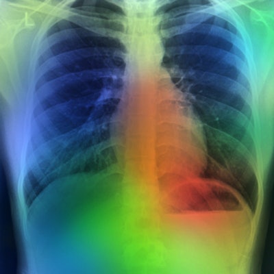
Now for the high-tech part. Artificial intelligence (AI) can be an effective way to address the challenging task of spotting subtle suspicious lung lesions on chest x-rays that could be a sign of COVID-19.
One such algorithm was described in a paper published July 21 in Radiology by a team from South Korea.
"Our findings suggest that a deep learning-based automatic detection algorithm (DLAD) may contribute to a reduction in the number of lung cancers overlooked on chest radiographs by improving observer performance," wrote a team led by Dr. Sowon Jang of Seoul National University Bundang Hospital in South Korea.
The authors determined that the AI algorithm improved the ability of readers to detect cancers that had been missed on chest x-ray. They also found that in healthy patients, there was no difference between the rate of chest CT recommendations when the algorithm was used and when it wasn't.
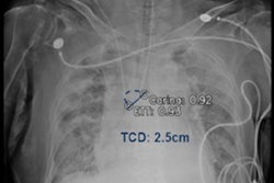
Another group developed an AI algorithm that can quantitatively assess the severity of COVID-19 on frontal chest x-rays, offering potential as a clinical triage and workflow optimization tool.
A team of researchers led by first author Dr. Matthew Li and senior author Jayashree Kalpathy-Cramer, PhD, from Harvard Medical School trained a special type of deep-learning algorithm called a convolutional Siamese neural network to provide a pulmonary x-ray severity score of patients with COVID-19. Their research was published online July 22 in Radiology: Artificial Intelligence.
In testing on internal and external datasets, the pulmonary x-ray severity scores generated by the algorithm correlated well with assessments by radiologists and could also help to predict if a patient would need intubation or would die within three days of admission.
Meanwhile, researchers at the University of South Florida and colleagues elsewhere utilized a readily available commercial platform to demonstrate the potential of AI to assist in the successful diagnosis of COVID-19 pneumonia on chest x-ray images, according to research posted May 26 in medRxiv.org.
The scientists investigated the potential for chest x-ray to be utilized along with AI to diagnose COVID-19 reliably. They wrote that the trained AI model overall demonstrated 92.9% sensitivity (recall) and positive predictive value (precision), with results for each label showing sensitivity and positive predictive value at 94.8% and 98.9% for COVID-19 pneumonia, 89% and 91.8% for non-COVID-19 pneumonia, and 95% and 88.8% for normal lungs.
They validated the program using chest x-rays of patients with confirmed COVID-19 diagnoses along with cases of non-COVID-19 pneumonia and normal x-rays. The model performed with 100% sensitivity, 95% specificity, 97% accuracy, 91% positive predictive value, and 100% negative predictive value, according to the scientists.
Turning to AI
And Mount Sinai Hospital in New York City also is turning to AI to help battle the pandemic. Research by Toussie has shown x-ray's value in helping physicians identify, triage, and treat high-risk patients.
"We are currently working to employ artificial intelligence to use the initial chest x-ray to predict outcomes, which would be incorporated into our electronic medical records system," she said in an email.
In addition, the University of Chicago Medicine has received a $20 million grant from the U.S. National Institute of Biomedical Imaging and Bioengineering (NIBIB) at the National Institutes of Health (NIH) to establish an extensive database of medical images from COVID-19 patients that researchers can use to better understand and fight the virus.
The project, called the Medical Imaging and Data Resource Center (MIDRC), will create an open-source database with medical images, including x-rays, from thousands of COVID-19 patients, the university said in a statement. By collecting and integrating images and their data using a dynamic, secure networked system, the MIDRC will provide a large-scale, open framework to enable technological advancements; guide researchers' validation and application of artificial intelligence tools; and translate clinical systems for the best patient management decisions.
University of Chicago will co-lead the center, along with an executive advisory committee that includes members of the American College of Radiology, the Radiological Society of North America, and the American Association of Physicists in Medicine.


