Researchers in South Korea have developed an AI model that can detect meningiomas on skull x-rays.
The approach involved integrating deep learning and traditional machine learning techniques, and could potentially level x-ray with advanced imaging modalities such as CT or MRI, noted Hyun Kim, PhD, of The Catholic University of Korea in Seoul, and colleagues.
“The proposed approach offers a promising diagnostic tool, especially for resource-limited settings where advanced imaging modalities might not be readily available,” the group wrote. The study was published November 17 in Scientific Reports.
Meningioma is the most common primary intracranial tumor, accounting for approximately 30% of all brain tumors. While the use of x-rays is limited for diagnosing meningiomas by their inability to detail soft tissue, they can reveal characteristic features such as thickening of the skull bone (hyperostosis), the authors explained.
The team explored the feasibility and efficacy of a hybrid AI approach designed to automatically detect these tumors on skull x-ray images.
First, the group used an image analysis model called EfficientNetB0 as the backbone and trained it on skull x-rays from 158 meningioma patients (632 images) and 201 control subjects (804 images) to focus on the areas where meningiomas resulted in bone changes. Next, they extracted these features from the images and fed them into traditional classifiers such as Random Forest and XGBoost, and then tested the model.
The internal test dataset consisted of 632 meningioma images and 804 control images (total 1,436). On these, the hybrid EfficientNetB0–Random Forest model achieved the highest accuracy (0.97) for detecting meningiomas, with an area under the receiver operating curve (AUC) of 0.999.
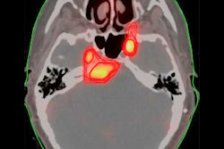
 Grad-CAM Visualizations for clinoid and tuberculum sellae meningiomas from the internal dataset. (a): Grad-CAM heatmaps for clinoid meningioma display dispersed activations, with reduced localization to the clinoid region, indicating limitations in explainability despite accurate model predictions. (b): For tuberculum sellae meningioma, activation maps show scattered and weakly localized activations, highlighting challenges in visual explainability for deeper anatomical regions.Scientific Reports
On an external dataset of skull x-rays from 103 meningioma patients and 103 normal skull x-rays, the Random Forest model achieved the best performance among classifiers, with an accuracy of 0.74 and an AUC of 0.76, the group reported.
Finally, Grad-CAM visualizations revealed that the model predominantly focused on specific regions of the cranial vault, particularly the frontal and parietal bones in cases of convexity and parasagittal meningiomas, which corresponded well with MRI findings.
“To the best of our knowledge, this is the first study to investigate the use of artificial intelligence with skull x-rays for early detection of meningiomas, suggesting a novel application of AI in enhancing diagnostic capabilities for this common intracranial tumor,” the researchers wrote.
They noted that this was a feasibility study, and future work will require validating the model on larger, more diverse datasets, with the goal of helping address the shortage of diagnostic experts and reducing healthcare costs.
The full study can be found here.