The amount of time a patient must remain motionless in a hospital bed after a coronary artery intervention can be reduced by more than half with the aid of a suture-mediated closure device, according to a paper in the latest issue of the Journal of Vascular and Interventional Radiology.
The prospective study, conducted by Dr. Denis Wetter and colleagues at the University Hospital Zurich in Switzerland, is one of a batch of recent papers that are praising the closure device, particularly when compared with tradition manual compression.
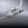
In the Swiss study, 100 patients underwent percutaneous transluminal coronary angioplasty (PTCA) between October 1997 and January 1998 and were randomized to a manual compression group or a suture-mediated closure group. In the latter, an over-the-wire suture-based vascular closure delivery system called Techstar (Perclose, Menlo Park, CA) was used. All patients received procedural bolus heparin during the intervention.
"Tissue approximation was achieved directly on top of the vessel wall through the channel of the puncture site, establishing immediate hemostasis…. Patients were kept in bed for approximately four hours until the first in-hospital follow-up," they wrote. Follow-up ultrasound imaging was done with a SonoDiagnost scanner (Philips Medical Systems, Best, the Netherlands) using a 7-MHz linear-array transducer for real-time and Doppler studies.
For patients in the suture-mediated closure group, the mean time to hemostasis was 7.3 minutes. Of the 50 patients, 30 were mobile after 4.2 hours; the remainder required more bed rest because of continuous oozing blood. However, all patients were discharged the next morning.
In the manual compression group, hemostasis occurred after 25.7 minutes. These patients also required more than 18 hours of bed rest after the procedure, 12 hours longer than most of the suture-mediated closure group.
In the closure study arm, Doppler ultrasound exams found three patients of the group of 50 had experienced hematomas ranging from 3 to 8 mL. In the manual compression group, imaging revealed that four patients out of 50 had developed 16 mL hematomas.
"A clinically occult small pseudoaneurysm (4 mL) was found on US in one patient who had undergone suture-mediated closure and in one patient (3 mL) who had undergone manual compression. No treatment was required; US examination at two-week follow-up showed spontaneous closure…. No patient in either group experienced major complications such as superficial infection or bleeding from the femoral access site," they wrote.
Based on these results, the researchers called suture-mediated closure a safe and effective method of achieving immediate hemostasis, as well as a good method for cutting down on the required period of bed rest. In addition, they found that ultrasound allowed them to catch more hematomas and study the impact of the closure on the arterial wall.
Other researchers have reported similar successful experiences with suture-mediated closures. The results of both arms of the Suture To Ambulate and Discharge Trial (STAND I and STAND II) came out in favor of suture-mediated closure. In STAND I, 200 patients underwent diagnostic procedures requiring cardiac catheterization. Using the closure device, hemostasis was achieved in 94% of the patients in a median of 13 minutes.
In STAND II, 515 patients were randomized to undergo either diagnostic or interventional procedures. Successful suture-mediated hemostasis occurred in 97% of the patients in a median time of 19 minutes with an ambulation time of 3.9 hours. In those patients who had manual compression, 98% achieved hemostasis, although it took 243 minutes and the patients were not ambulatory until almost 15 hours later (American Journal of Cardiology, April 1, 2000, Vol.85:7, pp.864-869).
In the Journal of Invasive Cardiology, a group from North Shore University Hospital in Manhasset, NY, detailed their clinical experience in four cases where suture-mediated closures were employed to manage an aortic balloon valvuloplasty site. The sutures were delivered prior to the intervention, using a 10-French closure device. The access site was then expanded to a 14-French size, facilitating adequate tissue capture, they said. The technique was successful in all four cases, and none of the patients suffered vascular complications (Sept. 2000, Vol.12:9, pp.474-477).
Finally, in that same issue of the Journal of Invasive Cardiology, a letter from Sanford Cardiology Associates in Sanford, FL, reported good results with a suture-mediated closure device to obtain hemostasis after a catheterization procedure performed via the brachial artery approach.
"Two successive catheterization procedures, using the right and left brachial arteries, were performed in a patient contraindicated for a procedure through the femoral approach," the Sanford group reported. "In both cases the closures were without complications. An aggressive anticoagulation regime could safely be prescribed to this patient because of the percutaneous surgic achievement of hemostasis," they wrote (Sept. 2000, Vol.12:9, pp.484-486).
By Shalmali Pal
AuntMinnie.com staff writer
October 4, 2000
Related Reading
Arterial puncture site management, July 2000
Let AuntMinnie.com know what you think about this story.
Copyright © 2000 AuntMinnie.com


