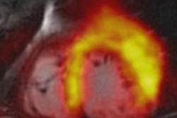
MIAMI BEACH, FL - In what is being billed as a first-of-its-kind study, researchers have concluded that simultaneous image acquisition by PET/MRI for myocardial perfusion imaging is feasible but "logistically demanding," according to a presentation on Sunday at the Society of Nuclear Medicine (SNM) annual meeting.
When patients were imaged at rest, researchers found no significant difference in performance between PET and MRI for myocardial perfusion measurement, either in total or by left ventricle segment. Under pharmacological stress, however, MRI underestimated myocardial perfusion in totality and by segment.
Lead study author Shelley Zhang from Technical University of Munich presented the results. She and her colleagues received one of six SNM Cardiovascular Council Young Investigator Awards for the research.
Gold standard
As Zhang noted in her talk, myocardial perfusion assessment can provide high diagnostic and prognostic information for the management of patients with coronary artery disease, but quantification of myocardial perfusion "remains a challenge" for the regional measurement of coronary flow reserve by a variety of imaging techniques.
Previous studies have established PET as the noninvasive gold standard for the quantification of myocardial perfusion under both rest and stress conditions, which could make recently developed integrated PET/MRI technology "uniquely suited to directly compare perfusion measurements in patients," she added.
The objectives of the study were to establish a protocol for the simultaneous acquisition of myocardial perfusion by PET and MRI, and to compare the simultaneous measurement of myocardial perfusion at rest and under pharmacological stress.
Researchers enrolled three male and two female patients with an average age of 63 years (± 13.8 years) in the study. Four patients had no known coronary artery disease, while one patient had a stent in the left anterior descending artery.
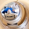
Patients were imaged with an integrated PET/MRI scanner (Biograph mMR, Siemens Healthcare), with two subjects asked to hold their breath during imaging and one person allowed to breathe freely. The PET device has a field-of-view of 25 cm and spatial resolution of 4.3 mm, while the 3-tesla MRI component included a six-element body surface coil and rigid spine coil.
'Precise planning'
The hybrid imaging protocol started with "precise planning," Zhang said. PET attenuation correction was obtained before injection of the MRI contrast agent gadopentetate dimeglumine (0.05 mmol/kg injected at 4 mL/sec). Multislice MR images were acquired at every heartbeat with an in-plane resolution of 2 mm to monitor the first-pass results from the contrast agent.
Arterial function was obtained using what Zhang described as "very short saturation delay time and very short echo time," followed by three high-resolution, short-axis MR images to give researchers single-heartbeat temporal resolution.
The five patients received an injection of adenosine for their stress test. Two minutes later, researchers started simultaneous acquisition of the gradient-recalled echo (GRE) MR image and PET image acquisition, immediately followed by gadolinium contrast for the MRI and ammonia for the PET image.
"For the resting scan, the same series of acquisitions were followed, except for the injection of adenosine," Zhang said. Finally, a late gadolinium enhancement measurement was performed.
In all, the researchers were able to acquire a total of 34 sectors in basal and middle ventricle slices for their analysis.
Heart rate
As for the results, the study showed a normal 30% increase in heart rate from 62.8 beats per minute at rest to 84.8 beats per minute at stress.
"For the left ventricular function in our population of patients, the ejection fractions and volumes are within the normal range," Zhang said. "Furthermore, the late gadolinium enhancement images did not show any sign of infarction."
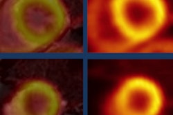
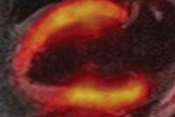
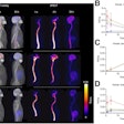
To correlate myocardial perfusion as assessed by PET and MRI, researchers first evaluated images from each patient's mid-left ventricle.
"We found an overall good correlation between [PET and MRI]," Zhang said. "Whereas resting flow agrees quite well, the stress values showed underestimation of MRI for [myocardial] perfusion."
After averaging the results from all sectors in three short-axis slices to obtain total myocardial perfusion values, the researchers again found "good agreement for the resting condition and an underestimation for stress conditions," she added.
'Logistically demanding'
Based on the results, Zhang and colleagues concluded "simultaneous-acquisition PET/MRI for myocardial perfusion imaging is feasible but logistically demanding."
While PET/MR imaging at rest showed "no significant difference" between the two modalities for myocardial perfusion measurement, either in total or by segment, myocardial perfusion calculated by MRI underestimated the results in total and by segment under pharmacological stress.
Still, Zhang added that integrated PET/MRI "offers a unique research tool to cross-validate physiologic measurements in humans."
The research was supported by the German research foundation Deutsche Forschungsgemeinschaft.

