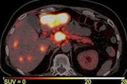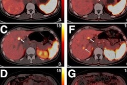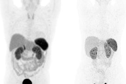
PET/CT imaging with the radiotracer gallium-68 (Ga-68) DOTATATE may reduce unnecessary biopsies in patients with neuroendocrine tumors (NETs) that have spread to the liver, according to research presented November 30 at RSNA 2022 in Chicago.
A group at Ludwig-Maximilians University in Munich, Germany, explored the diagnostic accuracy of the somatostatin receptor (SSR) radiotracer Ga-68 DOTATATE in PET/CT imaging for detecting liver metastases in differentiated NETs compared to histopathology. The imaging approach identified more cases than invasive biopsies, the team found.
"SSR-PET/CT can reduce unnecessary biopsies and optimize clinical patient management," presenter Dr. Matthias Fabritius told attendees.
Liver metastases in patients with NETs are an indicator for poor prognosis and significantly reduce patient survival. SSRs are proteins highly expressed on the cell surface of NET tumors, with radiotracers such as Ga-68 DOTATATE designed to bind to these targets and reveal tumor activity on PET imaging.
In a retrospective study, Fabritius and colleagues examined a total of 122 patients with differentiated NET (grade 1/2) and at least one suspicious SSR-PET/CT liver imaging finding with a histopathologic correlate. In total, 147 SSR-PET/CTs were evaluated independently by two readers. Histopathology was obtained by CT- or ultrasound-guided biopsy.
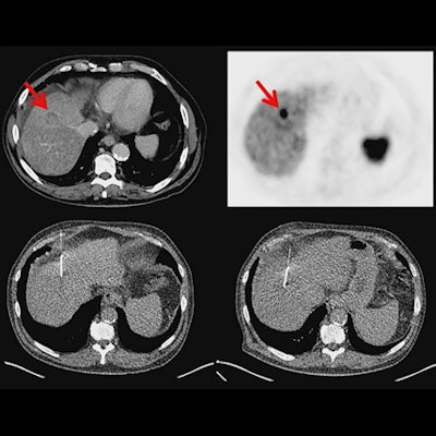
According to the results, SSR-positive metastatic hepatic involvement on PET/CT was reported in 98.6% (n = 145) cases, while histopathology showed hepatic involvement of NETs in 92.5% (n = 136) of cases. In 7.5% (n = 11) of cases, histopathology was negative, despite suspected liver metastases in SSR-PET/CT scans. In addition, in seven of 11 cases in which a second biopsy was available, SSR-PET/CT confirmed findings of liver metastases in five cases, while two of the rebiopsies confirmed the negative histopathologic results, Fabritius noted.
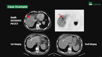 A case from the study showing uptake of the somatostatin receptor (SSR) radiotracer Ga-68 DOTATATE in a metastatic neuroendocrine liver tumor. Image courtesy of Dr. Matthias Fabritius.
Ultimately, the positive predictive value (PPV) of SSR-PET/CT for detecting liver metastases was excellent at 92.4%, but even higher at 95.9% when compared with histopathology and rebiopsy, according to the study.
Earlier in the session, Fabritius presented another study that showed SSR-PET/CT may also be comparable to contrast-enhanced liver MRI for evaluating liver metastases in NET patients, with the approach achieving a sensitivity of 97% and a specificity of 98%.
Both studies were retrospective and based on a large number of patients who underwent SSR-PET/CT at Ludwig-Maximilians University over a 15-year period. Research moving forward may combine these study designs for a more comprehensive picture, he said.
"SSR-PET/CT represents an important diagnostic tool in the detection of hepatic involvement in NET patients and should be part of NET tumor staging and restaging," Fabritius concluded.


