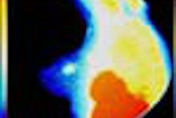
VIENNA – Italian researchers tested MRI and magnetic contrast echocardiography (MCE) to image patients with acute myocardial infarction and detect perfusion defects. They then compared the techniques to 99technetium sestamibi SPECT.
Dr. Luigi Natale from the Catholic University of Rome presented the results of the study Tuesday at the European Congress of Radiology. Fourteen consecutive patients with first acute MI (12 anterior, 2 inferior) underwent MR imaging, MCE, and SPECT within seven days after onset of symptoms.
MRI was performed on a short axis with multislice fast-spin echo and cine, single-slice first pass, and delayed T1-weighted fast gradient recalled echo (FGRE). The contrast agent used was 10 ml of Magnevist, injected intravenously at 3 ml/second. Segments were defined as Type 1 (normal first pass, delayed hyperenhancement); Type 2 (hypoenhancement at first pass, delayed hyperenhancement); and Type 3 (persistent hypoenhancement).
MCE was performed in harmonic power Doppler mode with Levovist given intravenously at 4 ml/minute. A perfusion defect was defined as absent or heterogenous enhancement, Natale said.
Finally, the SPECT scan was performed, and was used as the gold standard.
According to the results, MRI correctly identified the perfusion state in 75% of Type 1 segments. In Type 2 and 3 segments, MRI had a sensitivity of 79% and a specificity of 78% in comparison with SPECT. MCE diagnosed MI in 74% of the segments of infarct zones and correctly identified the perfusion state in 79% of the segments. Sensitivity was 79% and specificity was 78% in comparison with SPECT.
The group concluded that MCE is slightly superior to MRI in identifying perfusion defects, although MRI results may improve with a multislice first pace. Because of equipment limitations, this study was done with single-slice first pass, Natale explained.
"MCE loses some segments and this is the main problem with this technique," he said. "But we found quite a good concordance of 77% between MRI and MCE." By combining first pass and delayed MR imaging, the status of microvascular damage can be ascertained, he added.
In a second presentation, Natale used combined first pass and delayed contrast-enhanced MRI to assess myocardial viability after acute myocardial infarction (AMI).
In this study, a dozen consecutive patients with first AMI underwent MRI, 99Tc sestamibi SPECT and Dobutamine Echocardiography (DE) within one week after onset. Segments were classified as Type 1 (normal at first pass, absent or delayed enhancement); Type 2 (hypoenhancement at first pass, delayed hyperenhancement); and Type 3 (hypoenhancement at first pass and delayed imaging).
Types 2 and 3 were considered non-viable myocardia, while contractile reserve at DE and radionuclide uptake of greater than 50% were considered positive for viable tissue.
Using SPECT as the standard, Natale found that MRI had a sensitivity of 77% and a specificity of 100% for detecting viability.
By Shalmali PalAuntMinnie.com staff writer
March 9, 2001
Related Reading
SPECT identifies apoptosis in patients with acute MI, July 14, 2000
Angiography still best bet for heart attack patients, November 15, 2000
Click here to post your comments about this story. Please include the headline of the article in your message.
For more coverage of the European Congress of Radiology, visit our RadCast@ECR special edition.
Copyright © 2001 AuntMinnie.com


