MRI-based motion correction in simultaneous PET/MRI exams can significantly improve lesion contrast, signal-to-noise ratio, and image quality, compared with respiratory gating and no motion correction, according to a study published online June 28 in the Journal of Nuclear Medicine.
Researchers from Massachusetts General Hospital (MGH) believe they have not only confirmed the improvement in PET image quality in phantom and animal studies, but that their results also lay the foundation for the technique to be used effectively in simultaneous whole-body PET/MRI human studies.
A 'serious limitation' to PET
"Respiratory and cardiac motion is the most serious limitation to whole-body PET," according to the authors, as it causes fluctuations in attenuation measurements that can result in serious artifacts in reconstructed images (JNM, June 28, 2012).
With such motion corrected, simultaneous PET/MRI could provide enhanced lesion detection with no additional radiation dose to patients, wrote lead author Dr. Se Young Chun, from the Center for Advanced Radiological Sciences at MGH, and colleagues.
The researchers used a motion correction technique based on tagged MRI. The technique uses a combination of radiofrequency and field gradient pulses to create a magnetization pattern that persists over time and is distorted by motion, with the pattern moving with tissue. These distortions can be assessed on MR images.
All imaging studies were performed with an integrated PET/MRI scanner, with Siemens Healthcare's BrainPET unit inserted into the bore of the company's 3-tesla Tim Magnetom Trio MRI system. The PET/MRI system can be configured for animal studies. The inner and outer diameters of the PET gantry are 36 cm and 60 cm, respectively, and the axial and transaxial fields-of-view are 19.25 cm and 32 cm, respectively.
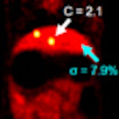
For the phantom study, the researchers built a phantom designed to mimic respiratory and cardiac motion. The device consisted of viscous methyl cellulose gel with background FDG activity (11.7 MBq), in which a balloon was suspended. Four spheres, each 1 cm in diameter, were attached to various locations with small and large motion amplitudes and patterns.
In the in vivo rabbit studies, six small lesions were prepared by soaking beads in an FDG solution and implanting them in the liver and diaphragm of the anesthetized rabbits. Three beads with 148 kBq to 222 kBq were placed in one rabbit, while three beads with 15 kBq to 56 kBq were placed in the second rabbit. The rabbits were also injected with 17.6 ± 1.8 MBq of FDG intravenously.
After one hour, simultaneous PET/MRI acquisitions were performed on each rabbit for 70 minutes. The second rabbit received an additional injection of 237 MBq of FDG at the end of the first acquisition sequence and was scanned for another two hours to obtain a second study with a lower lesion-to-background ratio.
In a primate study, Chun and colleagues anesthetized a monkey and injected it with 257 MBq of FDG. After two hours, simultaneous PET/MRI was performed for 30 minutes.
Motion 'virtually eliminated'
"Motion correction virtually eliminated motion blur without reducing [signal-to-noise ratio]," the authors concluded. The technique resulted in signal-to-noise ratios comparable to those obtained by gating with five- to eight-times longer acquisition times in all studies.
In the phantom study, the group found that MRI-based motion correction produced reconstructed images with fewer motion artifacts and less noise, compared with uncorrected or gated images. Lesion detection signal-to-noise ratio improved 166% to 276% with MRI-based motion correction versus gating. The authors noted a 43% to 92% improvement for the series of images with the largest motion displacement, compared with lesion detection without motion correction.
Similar results were seen in the animal and primate PET/MR images. The in vivo rabbit exams showed that MRI-based motion correction improved image quality to the same degree as in the phantom studies. "Our methods clearly improved the shape of the bead without increasing noise, compared with gating, which was comparable to the reference gated image," the authors wrote.
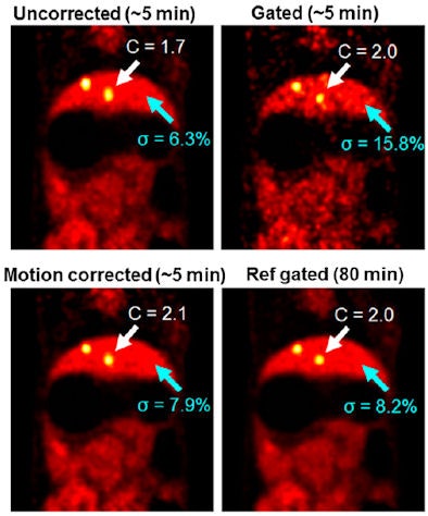 |
| Reconstructed PET images of a rabbit with uncorrected, gated, and MRI-based motion correction methods in the first scan. MRI-based motion correction reduced motion blur compared with the uncorrected image, without compromising noise as compared with the gated image. Images courtesy of JNM. |
In the primate study, the use of either gating or MRI motion correction improved image contrast in regions of interest by 240% to 280%. With gating, however, the improvement also resulted in 100% greater noise compared to the uncorrected images. "As expected, MRI motion correction yielded a level of noise similar to that of uncorrected images," the authors noted.
"To our knowledge, this is the first study to demonstrate the feasibility of in vivo MRI-based PET motion correction in simultaneous PET/MRI and the associated improvement in lesion detection" Chun and colleagues wrote. "The results of our studies can be considered a proof of principle, showing that simultaneous PET/MRI measurements can virtually eliminate respiratory motion blurring while improving the [signal-to-noise ratio] of the reconstructed images."
Extrapolating the results further, the researchers suggested that improved spatial resolution and lesion detection with MRI-based motion correction is feasible in clinical trials with no additional radiation dose to patients in simultaneous whole-body PET/MRI systems.


