Wednesday, December 4 | 11:00 a.m.-11:10 a.m. | SSK17-04 | Room S505AB
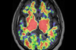
Despite differences in attenuation correction techniques, the PET images from a PET/MRI system are equivalent to PET/CT in terms of quality and detection rate for FDG-positive lung lesions, according to researchers from Germany.The study from Munich Technical University Hospital was led by Dr. Isabel Rauscher, a resident in the department of diagnostic and interventional radiology.
In the study, 40 patients underwent a dual-imaging protocol with FDG-PET/CT (Biograph 64, Siemens Healthcare) and PET/MRI (Biograph mMR, Siemens). Combined, both scans detected a total of 47 pulmonary lesions, with a mean size of 10 mm (± 11.4 mm) in 25 patients.
The PET datasets from both hybrid modalities detected 22 (47%) of the 47 pulmonary lesions with focal FDG update. Standardized uptake values (SUVs) of lung lesions in PET/MRI and PET/CT correlated significantly, with a tendency for higher SUVs in PET/MRI.
PET image quality was equivalent, and a high linear correlation coefficient in mean SUV for the PET images from PET/CT and PET/MRI was found, the group concluded.
"The detection rate of lung lesions can be significantly improved by adding a diagnostic contrast-enhanced VIBE [volumetric interpolated breath-hold examination] sequence to the PET/MRI protocol," the researchers added. "However, the detection rate of small lung lesions is still inferior compared to PET/CT with diagnostic CT of the chest."