Thursday, December 3 | 11:50 a.m.-12:00 p.m. | SSQ10-09 | Room E450B
With the help of texture analysis, MRI can distinguish between two uterine findings that have traditionally been difficult to separate, a group from Memorial Sloan Kettering Cancer Center reports.The study aimed to investigate whether qualitative MR features in combination with texture analysis can distinguish between atypical-appearing uterine leiomyomas and leiomyosarcomas.
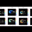
The researchers examined 41 women with either atypical-appearing leiomyomas or leiomyosarcomas, manually segmenting each tumor on T2-weighted axial MRI. They computed textures for each tumor with a variety of methods, recording the relationships between clinical characteristics, imaging features, and histopathology.
"We identified four qualitative MR features that accurately distinguish leiomyosarcomas from unusual leiomyomas, particularly if a lesion contains three or more of these MR features," lead investigator Dr. Yulia Lakhman told AuntMinnie.com.
Maximum sensitivity was about 95%, with specificity of 69%, according to Lakhman and colleagues.


.fFmgij6Hin.png?auto=compress%2Cformat&fit=crop&h=100&q=70&w=100)



.fFmgij6Hin.png?auto=compress%2Cformat&fit=crop&h=167&q=70&w=250)


