

A technique called saturation-transfer-based MR imaging shows promise for the evaluation of molecular changes and cerebrospinal fluid (CSF)-tissue water exchange in the brains of multiple sclerosis patients, researchers have reported.
The findings point to a protocol that could help clinicians better identify these changes, according to a team led by Ziyan Wang, PhD, of the University of Hong Kong in China. The results were published October 13 in the Journal of Magnetic Resonance Imaging.
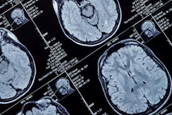
Multiple sclerosis is an autoimmune disease that attacks an individual's myelin, and MRI plays an important role in diagnosing and monitoring the condition, the group wrote. But "current standard MRI protocols for multiple sclerosis lack sequences capable of detecting molecular changes," it noted.
To explore an MRI framework that could identify these changes, the investigators used a technique called saturation-transfer-based MRI that consisted of chemical exchange saturation transfer (CEST) and magnetization transfer indirect spin labeling (MISL) sequences to quantify molecular changes and water exchange in the brains of multiple sclerosis patients. Their study included 52 participants, 21 of whom were multiple sclerosis patients and 31 of whom were healthy controls.
The MRI protocol was as follows: 3D inversion-prepared gradient echo T1w, 3D fast spin-echo T2w, 3D cube CEST ("cube" refers to replacing a slice-by-slice, plane-after-plane, 2D acquisition with a single 3D volume scan), and MISL on a 3-tesla scanner. The team used two methods to analyze the results: double-step multi-pool Lorentzian fitting (DMPLF) and Lorentzian difference analysis (LDA).
The group reported that the technique was effective for identifying changes in the brains of multiple sclerosis patients:
- CEST found decreased signals in the brain of multiple sclerosis patients using both DMPLF and LDA, with MPLF demonstrating superior performance in differentiating multiple sclerosis patients from healthy controls (AUC, 0.93).
- Multiple sclerosis patients showed a significant decrease (-6.6%) in brain tissue and an increase (+20.7%) in cerebrospinal fluid volume ratios compared to healthy controls.
"The saturation-transfer-based MRI framework can effectively evaluate molecular changes and CSF-tissue water exchange in the brains of multiple sclerosis patients," the authors concluded.
The complete study can be found here.

.fFmgij6Hin.png?auto=compress%2Cformat&fit=crop&h=100&q=70&w=100)


.fFmgij6Hin.png?auto=compress%2Cformat&fit=crop&h=167&q=70&w=250)


