Sunday, November 26 | 11:35 a.m.-11:45 a.m. | SSA17-06 | Room N228
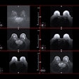
Researchers from Stanford University plan to provide additional evidence on the potential for ferumoxytol-enhanced MRI to detect and quantify macrophages in high-grade gliomas."Our study is a proof of concept for the clinical feasibility of visualizing and quantifying tumor macrophages in patients with malignant brain tumors using ferumoxytol-enhanced MRI," said study co-author and clinical assistant professor Dr. Michael Iv. "Results from our study show that ferumoxytol-enhanced MRI can be used as a noninvasive imaging biomarker of macrophages in adults with malignant brain tumors."
Iv and colleagues investigated eight patients with a mean age of 58.6 years and confirmed cases of high-grade glioma. Each patient received an intravenous infusion of ferumoxytol (5 mg/kg) with an MRI scan performed at least 16 hours later. The MRI protocol included quantitative susceptibility mapping (QSM) and R2* maps.
Patients' histopathology revealed iron particles only in CD68+/CD163+ macrophages. Macrophages are a key component of tumor-associated inflammation, angiogenesis, progression, and metastasis, as well as tumor response to therapy.
In analyzing the data, the researchers discovered statistically significant correlations between both QSM and R2* values and the number of iron-containing macrophages.
"This tool, which is widely available, can be used by radiologists and clinicians for predictive and prognostic purposes and can potentially offer improved monitoring and tracking of macrophage response of brain tumors to therapy," Iv told AuntMinnie.com. "Although this work specifically involves patients with brain tumors, potential applications of this work are diverse."
Other possible targets include cancers or other inflammatory-producing processes in and out of the central nervous system, as well as real-time tracking and improved delivery of stem cells or drugs paired with ferumoxytol across the blood-brain barrier.


.fFmgij6Hin.png?auto=compress%2Cformat&fit=crop&h=100&q=70&w=100)


.fFmgij6Hin.png?auto=compress%2Cformat&fit=crop&h=167&q=70&w=250)


