Wednesday, November 29 | 11:00 a.m.-11:10 a.m. | SSK09-04 | Room E450B
MRI's ability to detail changes in the prostate after focal ablation can help clinicians distinguish between normal findings and the possible recurrence of cancer, according to the findings of a study to be presented on Wednesday."We observed that appearance of the prostate after focal ablation changed over time, and these changes could be easily classified on MRI," lead author Dr. Andreas Hötker, currently in the radiology department at Universitätsmedizin Mainz, told AuntMinnie.com. Hötker led the research during his fellowship at Memorial Sloan Kettering Cancer Center in New York City.
More than 40 low- to intermediate-risk subjects with a mean age of 61 years underwent focal ablation therapy for prostate cancer between 2009 and 2014. Two radiologists reviewed more than 80 post-treatment MRI scans with the assignment to correlate the appearance of abnormalities relative to the number of days after focal ablation.
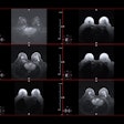
Within three weeks of the procedure, follow-up MR images revealed the presence of edema and rim enhancement of the ablation zone. Within eight weeks of focal ablation, the researchers discovered a hypointense rim around the ablation zone and the presence of an appreciable ablation cavity on T2-weighted MR images.
Some 14 months after focal ablation, follow-up MRI detected the formation of a T2-hypointense scar. One of the two radiologists also found the enhancement of the ablation zone and scar on follow-up MRI between 31 weeks and 85 weeks after the procedure.
"To effectively use MRI for follow-up after focal ablation of the prostate, radiologists must be familiar with the expected appearance of the prostate after ablation," Hötker noted. "Awareness of temporal patterns in the postablation appearance of the prostate should help radiologists distinguish normal MRI findings from possible recurrence and reduce image interpretation variability."


.fFmgij6Hin.png?auto=compress%2Cformat&fit=crop&h=100&q=70&w=100)



.fFmgij6Hin.png?auto=compress%2Cformat&fit=crop&h=167&q=70&w=250)


