Thursday, November 30 | 11:50 a.m.-12:00 p.m. | SSQ01-09 | Room E450A
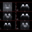
Japanese researchers plan to extol the diagnostic performance of texture analysis with dynamic contrast-enhanced breast MRI for nonmass enhancement and the differentiation of malignant and benign lesions.The MRI technique also appears valuable in distinguishing between ductal carcinoma in situ (DCIS) and inflammatory breast cancer.
"Extraction of quantitative MR imaging textural features from nonmass enhancement with various histology may be useful to classify the [nonmass-enhancing] lesions objectively," said study co-author Mariko Goto, PhD, an assistant professor in the department of radiology at Kyoto Prefectural University of Medicine.
Goto and colleagues reviewed 84 patients with 86 nonmass-enhancing lesions on breast MRI between March 2010 and March 2013. Textural features were taken from the lesions' volume of interest (VOI) as seen on both early and delayed phases of DCE-MRI performed on a 1.5-tesla system.
The researchers created a total of 138 textural features based on a volume of interest that included the contrast-enhanced area and a VOI that covered the entire region of enhanced and nonenhanced areas.
They found some statistically significant differences between the two VOIs regarding benign and malignant lesions, as well as DCIS and inflammatory breast cancer, but texture analysis was able to characterize the abnormalities regardless of the VOI parameters.
"Texture analysis of breast MRI for nonmass enhancement may be a promising tool to improve the diagnostic performance and may provide us with quantitative measures of the breast lesions," Goto told AuntMinnie.com.


.fFmgij6Hin.png?auto=compress%2Cformat&fit=crop&h=100&q=70&w=100)


.fFmgij6Hin.png?auto=compress%2Cformat&fit=crop&h=167&q=70&w=250)


