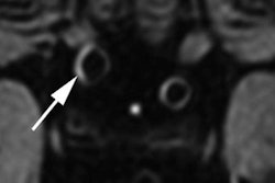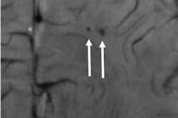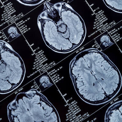
The ultrahigh field strength of 7-tesla MRI can identify brain abnormalities associated with epilepsy better than 1.5-tesla and 3-tesla MRI can, potentially changing the course of treatment for these patients, according to a study published online March 19 in PLOS One.
Researchers found epilepsy-related abnormalities in two-thirds of the subjects with epilepsy -- abnormalities not detected by lower-strength MRI scanners. The more powerful scanner also detected cortical lesions within the suspected epileptic seizure onset zone in 40% of the subjects.
Overall, the results from 7-tesla MRI prompted a change in surgical intervention and treatment planning for approximately 20% of patients.
"7-tesla MRI also revealed several subtle structural features in both patients and controls that were undetectable at lower field strengths, with significantly more abnormalities identified in epilepsy patients," wrote lead author Rebecca Feldman, PhD, and colleagues from the Icahn School of Medicine and Mount Sinai Hospital in New York City. "Therefore, information revealed by the 7-tesla exams has the potential to reveal biomarkers of epilepsy, provide enhanced lesion localization of focal epilepsy, increase the success of epilepsy surgery, and advance our understanding of the etiology of the disease."
While 1.5- and 3-tesla MRI scans are part of the routine workup before surgery for epilepsy, 20% to 30% of patients with focal epilepsy do not have a detectable lesion on exams at those magnet strengths. Such patients are less likely to be considered candidates for surgery, even though they possibly could benefit from such a procedure. In these situations, 7-tesla MRI has the potential to help.
To further investigate 7-tesla MRI's worth, the researchers enrolled 37 epilepsy patients (median age, 36 ± 14 years) and 21 healthy controls (median age, 34 ± 10 years). All of the subjects underwent diagnostic 1.5- or 3-tesla MRI scans based on a clinical protocol recommended for imaging epilepsy. They were then imaged on a 7-tesla whole-body scanner (Magnetom, Siemens Healthineers) with a 32-channel receive head coil.
The imaging protocol included magnetization-prepared rapid acquisition gradient-echo (MP-RAGE), MP2RAGE, T2-weighted turbo spin-echo (TSE), fluid-attenuated inversion recovery (FLAIR), and susceptibility weighted imaging (SWI) sequences.
Feldman and colleagues found 25 potentially epileptogenic abnormalities (67%) with 7-tesla MRI in the 37 patients with focal epilepsy; the abnormalities were not seen on lower-field MR images. The lesions were "definitely" or "likely" related to epilepsy in eight patients (21%). The detection of seven abnormalities (19%) directly changed the course of patients' surgical intervention and treatment planning.
Interestingly, the 7-tesla MR images showed structural and vascular abnormalities in all 37 epilepsy patients, as well as in 17 (81%) of the healthy controls.
The researchers used the high-resolution T2-weighted TSE images most frequently for their initial detection of subtle structural abnormalities, while the SWI sequence was "highly sensitive for the detection of focal susceptibility changes caused by or associated with cortical defects, cavernomas, and [developmental venous anomalies]," they wrote.


