When applied to 3D MRI, a new mapping algorithm can generate virtual placenta models that open up and flatten -- enhancing the visibility of the placenta during pregnancy, according to an upcoming presentation at the International Conference on Medical Image Computing and Computer Assisted Intervention in China.
The group of researchers, which includes computer scientists from the Massachusetts Institute of Technology (MIT), and a radiologist from Boston Children's Hospital, developed the volumetric mesh-based mapping algorithm to improve upon the visualization of placental anatomy and function.
Currently, physicians assess placental health during in vivo pregnancy monitoring using conventional ultrasound or MRI. However, these imaging modalities offer a limited supply of meaningful information due to the extensively curved surface of the placenta when still inside the uterus, the researchers noted.
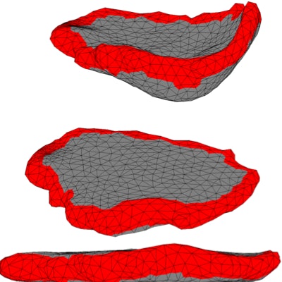
 A 3D placenta model based on MRI scans in an in utero position (top) and after flattening (bottom). Image and video courtesy of MIT CSAIL.
"The idea is to unfold the image of the placenta while it's in the body so it looks similar to how doctors are used to seeing it after delivery," lead study author Mazdak Abulnaga, a doctoral student from the MIT Computer Science and Artificial Intelligence Laboratory (CSAIL), said in a statement.
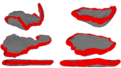
The algorithm works by employing matrix laboratory (Matlab, MathWorks) programming language to subdivide imaging data from a 3D MRI model into thousands of triangular pyramids, or tetrahedra. Then the algorithm rearranges the tetrahedra to resemble what it estimated the placenta would look like in a flattened position, as if the placenta were outside of the uterus.
In addition, the algorithm optimizes this transformed depiction of the placenta by balancing its estimate of the actual shape against image distortion. The algorithm's source code is available online via GitHub.
To validate their algorithm, Abulnaga and colleagues obtained 28 MRI scans from two prior pregnancy studies. They created 3D reconstructions of the scans and then applied their mapping algorithm to the resulting 3D MRI models.
Virtual depiction of a 3D MRI placenta model flattening.
They found the algorithm achieved subvoxel accuracy (i.e., less than a single 3D pixel) in mapping the boundary of the placenta, compared with a standard template. The 3D placenta models allowed the researchers to examine regions of the placenta that would have otherwise remained hidden on conventional MRI data.
For example, the models offered clear visualization of the placental cotyledons, structures that facilitate the exchange of nutrients between the mother and fetus. Being able to examine cotyledons in vivo could allow clinicians to diagnose and treat placental health complications much earlier in pregnancy and "help radiologists save time and more accurately locate problem areas without having to examine many different slices of the placenta," co-author Polina Golland, PhD, said.
"The biological processes that underlie cotyledon patterning are not completely understood, nor is it known whether a standard pattern should be expected for a given population," Chris Kroenke, PhD, of Oregon Health and Science University, said in a comment on the study. "This work will certainly aid researchers ... [and] provides a very elegant tool to address the issue of the placenta's irregular shape being difficult to image."
Looking ahead, the group plans to continue collaborating with Massachusetts General Hospital and Boston Children's Hospital on making the algorithm suitable for clinical use. They hope to conduct studies comparing the 3D placenta models in utero with images of the placenta after birth.
"While this is just a first step, we think an approach like this has the potential to become a standard imaging method for radiologists," Abulnaga said.


