Tuesday, December 3 | 10:40 a.m.-10:50 a.m. | SSG10-02 | Room N229
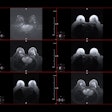
In this session, researchers will report on MRI's efficacy in assessing the dysfunction of blood vessels in the brain, or intracranial vasculopathies.A team led by Dr. Jae Song, an assistant professor at the Hospital of the University of Pennsylvania, searched databases through September 2018 for studies on intracranial vasculopathies that included vessel-wall MRI as the modality of choice. Among 2,431 articles, 79 met the inclusion criteria.
A majority of the published works came from Asia, and approximately two-thirds of the studies received federal funding. Intracranial atherosclerosis, which is the leading cause of stroke worldwide, was the most commonly studied intracranial vasculopathy.
Song and colleagues also found considerable heterogeneity in spatial resolution and magnet strength, which ranged from low-field 0.5-tesla scanners to 7-tesla systems. Postcontrast MR imaging was performed in approximately two-thirds of the cases surveyed.
The meta-analysis showed "considerable MR technical heterogeneity in MR imaging methods," the authors concluded in their abstract. Reducing the heterogeneity of MR protocols in vessel-wall MRI studies "should maximize effective synthesis and clinical translation of findings for intracranial vasculopathies," they added.


.fFmgij6Hin.png?auto=compress%2Cformat&fit=crop&h=100&q=70&w=100)


.fFmgij6Hin.png?auto=compress%2Cformat&fit=crop&h=167&q=70&w=250)


