NEW YORK CITY - Although still not as effective at correctly characterizing benign breast lesions, ultrasound elastography performs quite well in picking out malignancies, according to research presented at the American Institute of Ultrasound in Medicine (AIUM) meeting.
As part of their continuing evaluation of elastography, researchers from Elizabeth Wende Breast Care in Rochester, NY, found that the technique agreed with B-mode ultrasound on 97% of cancers. But elastography was only able to correctly point out 79% of benign lesions.
"In the clinical setting, [elastography] does prove to be an additional tool; I use it every day," Dr. Stamatia Destounis said. "It may be helpful to the breast clinician in the characterization of breast lesions."
Destounis presented the team's findings during a Friday scientific session.
Seeking to continue their evaluation of elastography in assessing breast lesions and its potential impact on biopsy rates, the Elizabeth Wende team enrolled 356 patients from their community-based breast center in their study. All patients had signed informed consent to receive elastography at the time of their ultrasound exam.
Patients received their ultrasound study on either a Sonoline Antares or S2000 scanner (Siemens Healthcare) with a 13.5 VFX or 14LB transducer. New solid lesions underwent needle core or fine-needle aspiration (FNA) biopsy.
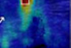
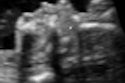
Images included standard B-mode ultrasound with and without size measurements and elastography with and without size measurements, Destounis said. The protocol comprised a dual-display cine clip and elastography color-mapping images.
The researchers recorded ratio measurements between B-mode and elastography, the size correlation between B-mode and elastography, and the likelihood of malignancy based on B-mode and elastography. Also noted were data on lesion laterality and location, as well as all imaging characteristics.
For lesions that were biopsied, the type of biopsy, biopsy results, and open surgical biopsy results were available. The researchers also had access to information on patient demographics, breast density, and follow-up visits.
The 365 patients yielded 371 elastograms and had a mean age of 49 (range 18 to 92). Lesion types included 200 masses, 107 fibroadenomas, 58 cysts, three architectural distortions, and one asymmetric calcification.
The average lesion size on B-mode was 1.4 cm (range: 0.3 to 4.6 cm) and 1.4 cm on elastography (range: 0.1 to 4.4 cm). Of the 220 biopsied lesions, 204 were masses, 12 were cysts, two were architectural distortions, and two were calcifications. Biopsy methods included 20 FNAs, 76 stereotactic biopsies, 112 with ultrasound, and 12 cyst aspirates.
In the 94 cancers in the study, B-mode and elastography had 97% agreement. The modalities differed in 1% of cases, and 2% of the cases were unclear.
Of the 122 benign cases, the techniques agreed in 79% of cases. They disagreed in 5% and the cases were unclear in 16%. There were also four atypical findings in which the modalities had 100% agreement.
"Our evaluation of elastography showed that it continues to be helpful in identifying cancerous lesions, identifying 97% of biopsied malignancies," Destounis said. "Elastography continues to be less effective at identifying benign lesions, at 79%."
Therefore, benign lesions on elastography require additional evaluation, she said.

