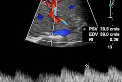
A new set of guidelines aims to standardize the terminology used to report arterial and venous spectral Doppler ultrasound waveforms. The document was jointly published on July 15 in Vascular Medicine and the Journal for Vascular Ultrasound.
The statement creates a designated set of key terms to describe findings on spectral Doppler ultrasound waveforms, the main diagnostic assessment for arterial and venous diseases. It was written by sonographers, vascular specialists, and other experts commissioned by the Society of Vascular Medicine and Society of Vascular Ultrasound.
"The hope of the writing committee is that this document will help us all to 'speak the same language,' and thereby advance the field of vascular ultrasound and improve patient care," stated lead study author Dr. Esther Kim, vascular labs medical director at Vanderbilt University Medical Center, in a press release.
The lack of shared nomenclature has been an ongoing problem for vascular ultrasound professionals. In fact, one out of five ultrasound professionals has had to perform a repeat arterial Doppler ultrasound examination because of terminology differences, according to a survey cited in the consensus statement.
"Over a decade ago, the lack of a standardized nomenclature to describe spectral Doppler waveforms was demonstrated to result in confusion amongst ultrasound professionals," Kim stated. "Not surprisingly, this can lead to negative clinical outcomes."
In the consensus statement, the committee established three major descriptors for ultrasound waveforms: flow direction, phasicity, and resistance for arterial waveforms and flow direction, flow pattern, and spontaneity for venous waveforms.
| Major descriptors for arterial ultrasound waveforms | |
| Flow direction | Antegrade
|
Retrograde
|
|
Bidirectional
|
|
Absent
|
|
| Phasicity | Multiphasic
|
Monophasic
|
|
| Resistance | High resistive
|
Intermediate resistive
|
|
Low resistive
|
|
| Major descriptors for venous ultrasound waveforms | |
| Flow direction | Antegrade
|
Retrograde
|
|
Absent
|
|
| Flow pattern | Respirophasic
|
Decreased
|
|
Pulsative
|
|
Continuous
|
|
Regurgitant
|
|
| Spontaneity | Spontaneous
|
Nonspontaneous
|
|
The statement also established terms that can be used to modify the main descriptors. For arterial waveforms, the seven modifying terms are as follows:
- Rapid upstroke -- Near vertical rise to peak systole
- Prolonged upstroke -- Abnormally gradual slope to peak systole; previously known as tardus, delayed, or damped upstroke
- Sharp peak -- Single, well-defined peak
- Spectral broadening -- Widening of the velocity band or filling in the typically clear area under the systolic peak; previously known as nonlaminar, turbulent, disordered, or chaotic
- Staccato -- High-resistance pattern with a short, low-amplitude diastolic signal punctuated by spikes of acceleration and deceleration
- Dampened -- Abnormal upstroke and peak, typically with decreased velocity; previously known as parvus et tardus, attenuated, or blunted
- Flow reversal -- Flow that changes direction but not as part of normal flow, can be transient or consistent with the cardiac cycle; previously known as pre-steal, competitive flow, or oscillating
For venous waveforms, the three modifying terms as follows:
- Augmentation -- Changes in flow velocity related to physical maneuvers, can be described as normal, reduced, or absent augmentation
- Reflux -- Persistent retrograde flow beyond normal closure time
- Fistula flow -- Flow with an arteriovenous fistula that becomes pulsatile due to communication with artery, sharp peaks often appear as pulsatile; previously known as arterialized or fistulous
In addition to creating the key descriptors and modifiers, the statement defined the reference baseline for spectral Doppler waveforms as the zero-flow baseline. It also advised against using the terms "normal" or "abnormal" to describe a waveform, since what is normal will depend on the part of the body and situation.
The statement also instructed sonographers to use image optimization techniques to acquire quality Doppler waveforms. This includes using an optimal transducer-to-vessel angle, the normal peripheral artery systolic waveform acceleration of 0.2 seconds, and proper transducer support.
Finally, the committee advised sonographers to provide complete descriptions for referring providers, including indication, relevant history, velocity measurements, and waveform characteristics. Sonographers should also include a conclusion with the clinical indication.
"We hope that this new Doppler waveform nomenclature will eliminate confusion and lead to appropriate diagnosis and better patient care," stated Dr. Raghu Kolluri, president of the Society of Vascular Medicine.


