Breast radiologists may want to consider the physical surroundings of mammography rooms for optimal image quality, according to a quality control study published November 15 in Radiography.
A team led by Stamatia Papathanasiou, PhD, from City University of London in England reported that low ambient light, a gray wall color, and monitors with high specification make for the best conditions for breast structure visibility. However, white wall color around monitors and high ambient light have negative impacts on image evaluation.
“Better standardization of the environmental condition is required in acquisition rooms,” the Papathanasiou team wrote. “Specifically, this research points to the benefit of using a low reflectance wall color and low illumination level around the monitors.”
Breast image interpretation requires radiologists to visualize structures on mammograms. Their respective performances are influenced by factors such as experience and fatigue. However, overall image quality also plays a role in accurate image interpretation, which is where the functional and environmental conditions of display monitors come into play. This includes display characteristics, illumination levels, and room design.
Papathanasiou and colleagues evaluated conditions that may impact quality control procedures within image acquisition rooms in breast imaging departments.
The study included nine test object images acquired from mammography phantoms. Sixteen observers evaluated the acquired images within 12 different environmental conditions, which included the following: low, medium, and high illumination level; white and gray wall color; and two monitors with high and low technical characteristics).
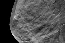
 The number of all visible structures was fewer at an ambient light of 500 lux compared to both 25 and 75 lux. In addition, structure visibility was better with a gray wall color at all ambient light levels. Paired comparison between monitors showed better structure visibility on the high specification monitor for all condition. Image is available for republishing under a Creative Commons agreement (CC BY-NC-ND 4.0).
The team reported the following findings:
- White wall color and the high ambient light level around the monitors had a statistically significant negative impact on the test object detectability. This included the number of multidirectional filaments, microcalcifications, and low-contrast detail discs visualized by the observers.
- The visibility of different structures was reduced at a high ambient light level (500 lux) compared to lower light levels (25 and 75 lux).
- The image with the most visible structures was obtained by the molybdenum/molybdenum (Mo/Mo) target filter combination and 20 mm polymethyl methacrylate thickness.
- Low-contrast discs in the upper part of the phantom image were less visible to observers. The researchers suggested that the direction and position of the light source that was used to control the ambient light in the room may have caused this.
The study authors further suggested that wall color's impact on mammogram structure visibility may be due to screen reflection.
“This work indicated the need for better standardization and optimization of the environment in acquisition rooms, to ensure consistency,” they added. “The lack of clear guidelines for acquisition rooms leads to a lack of standardization of the environment where the proper image evaluation and object detectability can be decreased significantly.”
The full study can be found here.


