How well does imaging predict residual tumor following neoadjuvant chemotherapy for breast cancer? It's a question mammographers at Massachusetts General Hospital in Boston are beginning to answer. In a presentation at the 2001 RSNA meeting in Chicago, Dr. Eren Yeh shared the preliminary results of her group’s study.
"Our purpose was to determine the relative value of mammography, ultrasound, and MR in predicting residual tumor as compared to the gold standard of physical examination and pathology," Yeh said. "More frequently, patients with large breast cancers are getting neoadjuvant chemotherapy."
One of the advantages of this treatment is that the tumor mass is decreased, thereby downstaging large cancers. In addition, neoadjuvant chemotherapy can treat micrometastases, and, because the tumor is still intact, in-vivo assessment of the therapeutic benefits is possible, Yeh added.
Twenty-five patients with palpable breast tumors have been enrolled to date for this prospective study. At the initial diagnosis, 20 patients had invasive ductal cancer, four had invasive ductal cancer plus ductal carcinoma in situ (DCIS), and one patient had invasive lobular cancer.
Imaging was used to gauge the effects of sequential single-agent chemotherapy (adriamycin followed by taxol or vice versa) on physiologic parameters and molecular markers. All patients underwent MR and ultrasound exams and mammography prior to, and following, treatment. Dynamic high-resolution MRI was done on both breasts on a Signa 1.5-tesla system (GE Medical Systems, Waukesha, WI).
As of November 2001, 11 patients had completed the treatment protocol. The imaging studies were reviewed by two radiologists and compared to clinical response and pathology.
According to the results, a partial clinical response was seen in 10 of 11 patients. One patient had stable disease upon examination. The response agreement for MRI was 65% when compared with physical exam. For ultrasound, the response agreement was 53%; for mammography, 41%.
The response agreement between pathology and imaging was as follows: MRI, 71%; ultrasound, 41%; and mammography, 24%.
"One of the reasons we think that mammography, as compared with pathology, didn’t do as well is because a lot of our patients had dense breast tissue," Yeh explained. "Eighteen of 25 patients had an ACR parenchymal pattern of 3 or 4. In six of our patients, the tumor mass was not visible at all on mammography, although some did have associated calcifications, which were visible."
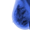
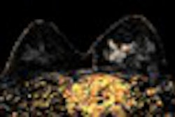
In one case, a 46-year-old patient had a lump in her left breast. Prior to chemotherapy, no discreet masses were identified on mammography; on ultrasound, there was an ill-defined hypoechogenic shadow in the mass. After chemotherapy, the tumor mass was smaller in size based on post-treatment ultrasound. The pre-chemo MR showed extensive linear branching enhancement, which turned out to be much more extensive than originally seen on ultrasonography, mammography, or with physical exam. Post-treatment, there was complete resolution of enhancement on MR, Yeh said.
"As we were looking at these MRIs, we noticed that they fell into different enhancement patterns," Yeh said. "(Eight patients had) masses with the defined margin. Four patients had multiple nodules. Seven patients had a complex diffuse tissue infiltration type of enhancement, and four patients had linear branching."
In six patients who had undergone complete surgical excision of the tumor, MR was the most accurate in predicting residual tumor in four of those patients. In the other two, the modality overestimated the amount of residual disease when compared with pathology.
"In conclusion, MRI appears to provide the best correlation with pathology, better than physical exam, mammography, and ultrasound in patients undergoing neoadjuvant chemotherapy. However, MRI did overestimate residual disease in a small percentage of the patients," Yeh said.
By Shalmali PalAuntMinnie.com staff writer
January 7, 2002
Related Reading
Patients with known breast malignancy benefit from MRI, November 25, 2001
MRI superior to mammography in screening high-risk women for breast cancer, July 27, 2001
Software pairs MRI and mammography to detect subtle pathology, April 27, 2001
A year after breast conservation therapy, MRI shows high diagnostic confidence, January 26, 2001
Copyright © 2002 AuntMinnie.com


