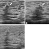
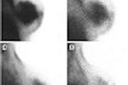
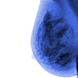
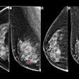
A real-time, handheld near-infrared device could offer a simple and noninvasive way to conduct breast cancer imaging, according to researchers from the University of Pennsylvania in Philadelphia. Using diffuse optical tomography, the device relies on near-infrared light to image areas of the body and create spatially resolved maps of their optical properties.
Dr. Saroja Adusumilli discussed the results of a pilot study that used the device at the 2002 American Roentgen Ray Society meeting in Atlanta. "This technology functions well as a medical probe because light is nonionizing over many wavelengths, and red, and near infrared, light actually passes easily through gross tissue," Adusumilli explained.
"The breast is also a very good model because it’s on the surface of the body, so it’s accessible to instruments," she said. "Breasts have a lot of fat and lipids and it is a relatively low scattering and attenuating medium."
A light source in the 700-800-nanometer range can provide enough light to penetrate tissue from 2-12 cm. In this range of wavelengths, oxygenated and deoxygenated hemoglobin are the main absorbers of light. By measuring their scattering and absorption coefficients, two phenomenon of tumor development can be exploited, she added.
"The first is angiogenesis. The development of new vessels around a lesion is considered to be a critical event that takes a cluster of barren cells and turns it into a rapidly growing malignancy. We hypothesized that measuring elevated blood volume in a lesion relative to the normal tissue around it can imply the presence of increased vascularity," she said.
The second phenomenon is hypermetabolism, or an imbalance between oxygen delivery and uptake. In tumors, uptake generally exceeds delivery, resulting in tissue hypoxia and areas of tumor necrosis.
For the study, 27 women with either a palpable mass or an abnormal mammogram underwent optical imaging, while 34 healthy patients formed a control group. Histopathology was used as the gold standard.
The handheld imager consists of a central light source that is made up of three light-emitting diodes, all with three distinct wavelengths: 730, 805, and 850 nanometers. These diode detectors estimated tumor blood volume and deoxygenated hemoglobin concentration.
"There are eight silicone diode detectors that are placed in the periphery, about 4 cm from the center (of the device). And these detectors are mounted on a sponge rubber pad, covered by plastic wrap. This is the pad that’s ultimately placed on the patient’s breast. This device is also hooked up to a power supply and to a laptop computer that collects the data," Adusumilli said.
The device was placed on a portion of the breast that was known to have a lump based on ultrasound or mammography results.
"We usually place Detector 1 over the lesion. Then the other set of detectors is placed over relatively normal breast tissue. Really, no breast compression is involved aside from the pressure that the operator is placing to hold on to the device," she said.
The light source was activated and data acquired over 1-8 seconds. The device was rotated so that each of the eight detectors would be placed on the lesion.
"This gives us more data points, and it also minimizes the influence of any potential for any variation that the operator might be creating by changing the pressure in the placing of the device," Adusumilli said. "We then performed the same procedure in a mirror-image location of the contralateral normal breast as a control."
According to the study results, 15 out of 31 lesions were found to be cancerous on pathology, with lesions ranging in size from 4 mm to 4 cm.
"The remaining lesions were benign. We found that blood volume, which is a marker for angiogenesis, and deoxygenated hemoglobin concentration, which is a marker for hypermetabolism, were elevated in malignant tumors relative to normal breast tissue and to benign lesions," such as cysts and fibroadenomas, Adusumilli said.
Based on spatial congruence and a 2-D nomogram of blood volume versus deoxygenated hemoglobin, the sensitivity of the infrared imager was 92%, the specificity was 100%, and the positive predictive value was 100%.
"These preliminary results suggest that this device does have the potential to detect the cancer signatures of a malignant lesion by looking at markers of angiogensis and hypermetabolism," she said.
The method does have some limitations at present: The infrared imager can study depths up to 2 cm. As a result, it may miss small lesions in a large breast or deep within the tissue. Also, the technique is operator-dependent -- an unskilled operator could alter the data points.
Finally, the pilot study consisted of a small group of patients. A larger patient population will have to be looked at in order to determine whether infrared imaging can be used as a complement to conventional breast imaging, she said.
By Shalmali PalAuntMinnie.com staff writer
July 19, 2002
Related Reading
Tiny US lens-transducer gauges intra-arterial turbulence, April 25, 2002
Near-infrared spectroscopy can identify plaques likely to rupture, April 24, 2002
Investigational infrared technology may help identify stent restenosis risk factors, May 4, 2001
Researchers join race to develop optical breast imaging device, April 27, 2001
Copyright © 2002 AuntMinnie.com


