Back in March, the American Cancer Society (ACS) lent its qualified support to the idea of screening women at high risk of breast cancer with MRI, helping to cement that modality's role in monitoring this younger patient population. A new study in Radiology puts MRI to the test, and up against ultrasound, for the surveillance of women at high risk.
"Genetic predisposition accounts for an estimated 5%-10% of all breast cancers," wrote Dr. Constance Lehman, Ph.D., and colleagues. "The American Cancer Society currently suggests that women discuss with their clinicians the potential benefits and risks of adding alternative screening methods such as US or MR … few prior studies have included both US and MR for the detection of clinically and mammographically occult cancer in women at high risk" (Radiology, August 2007, Vol. 244:2 pp. 381-388).
Lehman lead the International Breast MRI Consortium (MBC) and the Cancer Genetics Network (CGN) for this prospective analysis, done on 171 women (mean age of 46), 91 of whom had undergone genetic testing for familial cancer risk. Eligibility criteria included the presence of BRCA mutations (73 of 91 deemed carriers), family history, and Ashkenazi Jewish ancestry.
All patients underwent clinical breast exam, mammography, sonography, and MRI within 90 days of each other. Ultrasound was done first using an 8-MHz or greater transducer, with the entire breast scanned in two planes and coded according to the BI-RADS lexicon for ultrasound. Two-view mammographic study of each breast was performed and coded according to BI-RADS lexicon.
MR studies were conducted on a 1.5-tesla magnet with a dedicated breast coil. The protocol included fast-spin echo (FSE) images with fat suppression and T1-weighted images acquired before and after the injection of 0.1 mmol/kg of gadolinium. MR images were assessed on a five-point scale with 1 representing a negative test and 5 considered highly suggestive of malignancy.
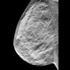
Fifteen readers participated, with separate readers assigned for each examination. According to the results, over 65% of the women had heterogeneously or extremely dense breasts. Among the 171 subjects, six infiltrating ductal carcinomas were detected: two with mammography, one with ultrasound, and all six with MRI. Of these six cancers, half were in women with heterogeneously dense breast tissue.
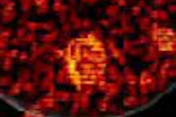
While 100% of the cancers were detected with MRI, the authors pointed out that MR exams did lead to more patients being recalled for additional imaging or biopsy (24%). The positive predictive value (PPV) of biopsies based on MR results was 43%. In comparison, based on ultrasound results alone, 9% of the patients were recalled and the PPV for biopsy was 25%.
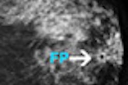
"The added cancer yield was associated with a higher biopsy rate with MR (8.2%) compared with … US (2.3%) imaging," Lehman's group wrote. "MR had a higher sensitivity in cancer detection at the cost of higher biopsy rate. In our study, US did not identify cancers missed at mammography."
The researchers offered some possible reasons why ultrasound did not perform as well. First, previous ultrasound trials that looked at mammographically occult cancers did not include MR, so there was no reference standard for comparison. In addition, breast density presents a greater challenge to sonography than MR.
The group acknowledged that their patient population was small, and that the study consisted of a single round of screening without long-term follow-up. Nevertheless, they concluded that MR is an "important complement to mammography" in high-risk women, giving it an edge over ultrasound.
By Shalmali PalAuntMinnie.com staff writer
August 6, 2007
Related Reading
Breast MRI helps guide surgical treatment, May 22, 2007
MRI becoming more efficient in breast cancer detection, May 18, 2007
Mammography still offers advantages over breast MR in DCIS, April 20, 2007
MRI useful for detecting cancer in contralateral breast, March 28, 2007
ACS advocates breast MR screening for high-risk women, March 28, 2007
Copyright © 2007 AuntMinnie.com


