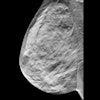

Quantification of breast density could identify women at increased risk of breast cancer who might benefit from more frequent screening and/or an alternative imaging examination, such as breast MRI or ultrasound. It would also provide a more precise assessment of the dose absorbed by the breast during mammography.
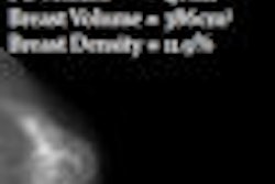
With these applications in mind, researchers from the University of Toronto in Ontario and the University of California, Davis have shown how measurements of volumetric breast density can be obtained from digital mammograms (Physics in Medicine and Biology, June 7, 2010, Vol. 55:11, pp. 3027-3044). The method builds upon previous work that allows the breast thickness to be determined precisely -- a key challenge when working in 3D.
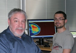 |
| Martin Yaffe (left) and PhD candidate Olivier Alonzo-Proulx (right). The computer screen shows an image of the volumetric density from a mammogram. |
The algorithm proposed in the paper by lead author Olivier Alonzo-Proulx, a doctoral candidate in the University of Toronto's department of medical biophysics, and colleagues builds on an earlier method for measuring breast density in which the transmitted signal measured on breast tissue-equivalent calibration objects was related to their known thickness and compositions. This refined algorithm, Cumulus V, has been adapted to digital mammography and uses a combination of measurement and modeling to estimate the compressed breast thickness.
To validate Cumulus V, the researchers applied the algorithm to a series of simulated mammograms, derived directly from volumetric images acquired on a dedicated 3D breast CT scanner. This involved applying a finite element model to the CT images to simulate the mechanical breast compression of a mammography examination. A physics model of a digital mammography system was then used to simulate the transmission of x-rays through the deformed breast volume.
"The algorithm performed well," Yaffe told medicalphysicsweb. "The relation between the CT data and our algorithm was almost one-to-one, and the difference between the two varied between two and three percentage points."
One potential weakness with the validation strategy, which relied on simulated mammograms, was that the results were affected by inaccuracies in the physics model of the imaging system. "But we were able to estimate the errors due to the model, which were small and comparable to the errors due to the algorithm only," Yaffe said. "Another approach would be to test the algorithm on mammograms of patients who have also undergone CT imaging."
Cumulus V will now be applied to the "thousands" of digital mammograms obtained in the various regional hospitals with which the researchers are affiliated. A case-control study is planned that will relate volumetric breast density to parameters including cancer occurrence, age, and breast volume. A study group will also be formed to look into the effects of breast composition on absorbed dose.
In addition, the breast density measurements are to be incorporated into a multiparameter model for risk of breast cancer that combines volumetric density with other factors, such as family history and body mass index.
By Paula Gould
Medicalphysicsweb contributing editor
July 2, 2010
Related Reading
Mātakina tackles breast density problem with Volpara software, February 3, 2010
© IOP Publishing Limited. Republished with permission from medicalphysicsweb, a community Web site covering fundamental research and emerging technologies in medical imaging and radiation therapy.

