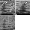
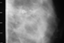
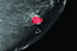
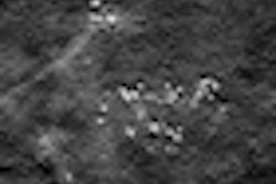
Screening mammography finds smaller breast tumors with less nodal involvement than clinical exams in women 40 to 49 years of age, and women whose tumors were detected by mammography had higher survival rates compared to those with tumors detected by clinical exam, according to research presented this week at the American Society of Breast Surgeons (ASBS) annual meeting.
The findings indicate that excluding this population from annual screening exams under the revised U.S. Preventive Services Task Force (USPSTF) 2009 mammography guidelines would negatively affect women's survival, the researchers believe.
Lead researcher Dr. Paul Dale at the University of Missouri-Columbia and colleagues conducted a 10-year retrospective study that looked at 1,581 breast cancer patients treated between 1998 and 2008. Of the patient cohort, 20% were between the ages of 40 and 49; 47% of patients were diagnosed through mammography and 53% through clinical exams or other nonmammographic methods.
In the mammography group, the mean tumor size was 20 mm in diameter, while nonmammographically identified tumors had a mean size of 30 mm. The study also found that the frequency of lymph node involvement in the clinically detected patients was approximately twice that of the mammographically detected patients.
Using survival analysis methodology, the team estimated the study participants' five-year disease-free survival rate to be 94% for women whose cancer had been identified with mammography, compared with 78% for those whose cancer had been identified clinically, according to Dale.

