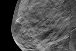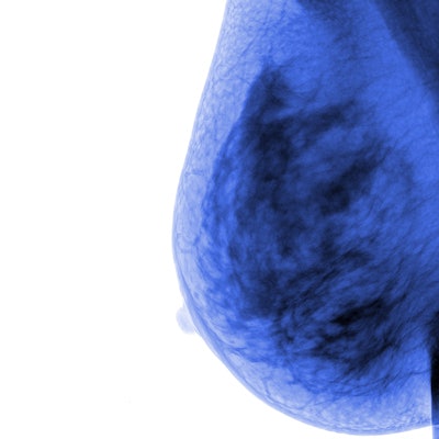
Digital breast tomosynthesis (DBT) finds 13% more invasive and early-stage cancers in women with dense tissue than conventional 2D mammography, according to a study published in the January issue of the Korean Journal of Radiology.
The study results strengthen DBT's place in the breast cancer screening arsenal for women with dense tissue, wrote a team led by co-first authors Dr. Soo Hyun Lee and Dr. Mi Jung Jang of Seoul National University Bundang Hospital.
"Our findings indicated that DBT images were superior to conventional [2D mammography] images with regard to breast cancer detectability in dense breasts," the group wrote.
The researchers compared the performance of DBT and 2D mammography in identifying breast cancer in patients with dense tissue. Five radiologists used a scoring scale of 0 to 2 to assess the detectability of 300 breast cancers in 288 women with dense tissue on DBT and 2D images. Each woman also underwent breast MRI to track the location and size of cancer lesions (Korean J Radiol, January 2019, Vol. 20:1, pp. 58-68).
The researchers also recorded the women's hormone status, histologic grade, and breast cancer subtype (human epidermal growth factor receptor 2, or HER2) to explore which factors might influence the performance of the two modalities. Most women (66.3%) had heterogeneously dense tissue; 33.7% had extremely dense tissue.
Lee, Jang, and colleagues compared the following categories:
- Group 1: DBT-only detected cancers
- Group 2: Cancers found on both DBT and 2D mammography
- Group 3: Cancers missed by both modalities but identified on MRI
The researchers found that 40 cancers (13.3%) were identifiable only on DBT, 191 (63.7%) were detectable on both DBT and 2D mammography, and 69 (23%) weren't found by either modality. Cancer detectability scores were higher for DBT than for 2D mammography (p < 0.001), and the cancers found only on DBT were more likely to be invasive, lobular breast cancers. DBT was less effective in identifying ductal carcinoma in situ (DCIS).
| DBT, 2D mammography, and breast MRI for identifying breast cancer in dense tissue | |||||
| Performance measure | Group 1 (DBT only) | Group 2 (DBT + 2D mammo) | Group 3 (MRI) | p-value, groups 1 and 2 | p-value, groups 1 and 3 |
| Total cancers | 13.3% | 63.7% | 23% | -- | -- |
| Invasive ductal cancers | 70% | 70.4% | 60.9% | 0.002 | 0.004 |
| DCIS | 5% | 16.8% | 27.5% | 0.002 | 0.004 |
| Low-grade cancers | 22.5% | 10% | 33.3% | 0.026 | 0.490 |
| HER2-negative cancers | 92.3% | 70.5% | 87.3% | 0.004 | 0.525 |
"Forty breast cancers (13.3%) were more detectable on DBT and the detectability score was higher on DBT than on conventional [2D mammography]," the researchers wrote. "Our result of [a] 13.3% increase in cancer detection on DBT is comparable to those of single- or multicenter studies, which reported a 10% to 51% increase in cancer detection."
The findings suggest that DBT may be a better choice than 2D mammography for screening women with dense tissue, the investigators concluded.


