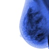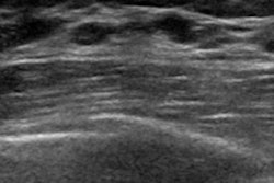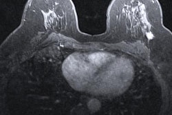
MRI and contrast-enhanced digital mammography (CEDM) are more accurate in judging the final size of breast tumors after endocrine therapy, according to research to be presented April 8 at the American Society of Breast Surgeons (ASBS) annual meeting in Las Vegas.
In her presentation, Dr. Chi Zhang from the Mayo Clinic in Scottsdale, AZ talked about her team's findings, which compared four different imaging modalities when it comes to measuring residual in-breast disease after therapy.
"Imaging is important across the board for breast cancer," Zhang told AuntMinnie.com. "It's how we plan and stage cancers through the different types of imaging available to us."
Medical imaging helps with monitoring tumor response to breast cancer therapies and can influence decision-making between breast conservation and mastectomy. However, there is no uniform protocol when it comes to women receiving neoadjuvant endocrine therapy.
Zhang and colleagues wanted to find how accurate different imaging modalities are in predicting breast tumor response in patients who underwent neoadjuvant endocrine therapy, using CEDM, MRI, conventional mammography, and ultrasound.
"Because imaging is such an important part of managing breast cancer, we really want to make sure we can track the progress of disease while patients are on therapy and make surgical decisions," Zhang said.
The researchers looked at data from 119 women with an average age of 58.7 years. The women had confirmed invasive breast cancer and underwent at least three months of neoadjuvant endocrine therapy, as well as post-treatment imaging between 2013 and 2021.
| Comparison of imaging modalities in measuring breast tumor size post-neoadjuvant endocrine therapy | ||||
| Conventional mammography | Ultrasound | CEDM | MRI | |
| Accuracy frequency (within 1 cm) | 48.1% | 54% | 57.5% | 58.3% |
| Agreement between imaging findings and pathology size | 0.16 | 0.24 | 0.38 | 0.45 |
| Correlation to pathology size | r = 0.21 | r = 0.42 | r = 0.49 | r = 0.52 |
Zhang said that one possible strategy for clinicians is to use ultrasound as a first line of imaging while patients are undergoing therapy and then use contrast-enhanced digital mammography for final surgical planning. This is because ultrasound is easier to access and is more cost-efficient. Ultrasound also showed moderate correlation with tumor size on pathology.
Zhang also said she was surprised conventional mammography lagged further behind the other imaging modalities.
Future research is not currently planned in relation to this study. However, Zhang told AuntMinnie.com she would be interested in exploring how neoadjuvant endocrine therapy can impact costs and quality of life for women.
"In the past, we've shown that smaller surgery and preserving the natural breast parenchyma does lead to improved quality of life in that women are more satisfied with their physical appearance," she said. "My thought is that less radical surgical intervention can reduce perioperative morbidity and preserve quality of life and optimize cosmetic outcomes down the road for women."


