INDIANAPOLIS - Contrast-enhanced mammography (CEM) continues to show potential, but implementing this modality into breast imaging departments takes several steps, according to a July 11 presentation at the AHRA annual meeting.
In her talk, LaJuana Fuller, director of women's imaging at the University of Pittsburgh Medical Center (UPMC)-Magee Womens Hospital, discussed how the hospital's breast imaging department implemented its CEM protocol, as well as its challenges and opportunities.
"I believe that the future looks bright for CEM," Fuller said. "As research continues to take place, it'll show where the benefits are and who will be eligible. I think it's worth considering for your organization."
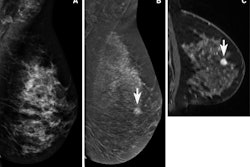
CEM in research has gained traction in diagnostic breast imaging as a possible alternative to supplemental MRI. Some benefits highlighted in research include not being exposed to loud noise or claustrophobic conditions experienced by some patients undergoing MRI, as well as patients being able to lie in the prone position.
 LaJuana Fuller discusses at the 2023 AHRA annual meeting how UPMC-Magee Womens Hospital implemented the medical center's contrast-enhanced mammography program. She said it involved stakeholder engagement, staff training, and communication. She said the future is bright for the modality as more studies highlight its potential benefits in diagnostic breast imaging.
A recent study also led by UPMC researchers found that women overall prefer undergoing CEM over MRI for the above reasons along with others.
Fuller shared her experience with UPMC implementing its CEM program, in which talks began in 2014.
"We're fortunate that we have a very engaged staff," she said. "This was taking us into an unknown kind of workflow."
Equipment and staff training
Fuller said departments need to make sure they have the following equipment for successful CEM implementation: a mammography unit that has CEM software compatibility, a CT power injector, contrast media, point-of-care blood analysis, and intravenous (IV) supplies.
When UPMC's CEM program was first developed, a half-day training session was held, with volunteers from each health center within UPMC attending. After completing the session, the volunteers were evaluated for competency and completed a course on contrast reactions.
With this new equipment comes new training for staff members, which Fuller said means a cultural shift in the breast imaging department.
"When we first started, some people weren't happy," she said. "That's part of the work as an administrator, to shift that culture and to communicate why this is happening."
Involving stakeholders and communication
When Fuller was introduced to the concept of CEM, she reached out to one of her nurse leaders in the education department. From there, they developed their training program.
Fuller said it's important to involve stakeholders such as finance and administrative teams among others into discussions about planning and implementing CEM protocols and acquiring the necessary equipment. She added that medical stakeholders such as technologists, referring physicians, and surgeons should also be involved.
"You're adding a new service, so want them to be at the table too," she said. "You want them to endorse what it is you're bringing into your breast imaging program."
Along with that, she said it's important to work with marketing communications teams to effectively communicate with community members and potential patients in language they can understand.
Prescreening patients and the protocol
While CEM has its share of benefits, patients must be prescreened before undergoing this exam. Fuller said that currently, CEM is offered at Magee Womens Hospital when patients face contraindications for breast MRI.
Women who have contrast allergies, have kidney issues, or use certain medications may not be allowed to undergo a CEM exam.
Fuller and colleagues found that CEM exam times are shorter than MRI exams. She said imaging should be performed about 2.5 minutes after contrast injection, with craniocaudal views taken first and mediolateral oblique views second. Additionally, she said the affected breast should be imaged first. The imaging process takes about seven minutes, with turnaround time being around three days on average.
Bright future
Fuller said that today, CEM is used in five centers at UPMC. She owed its successful implementation to a strong leadership team.
Along with the aforementioned UPMC study on patient perceptions, researchers from the medical center are also leading studies on CEM vs. MRI on overall performance in detecting cancer.
"We're constantly moving and evolving our technology to improve detection of breast cancer," Fuller said. "One thing that's unique ... is that we're always looking for what we can do more of, not just with technology but also in the community with addressing disparities and closing gaps ... so that patients' family members can be with them."


