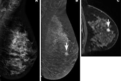
Contrast-enhanced mammography (CEM) may help accurately downgrade BI-RADS 4 breast lesions, a study published August 23 in the European Journal of Radiology has found.
A team led by Anna Grażyńska, MD, from the Medical University of Silesia in Katowice reported that CEM had high negative predictive value (NPV) when it came to assessing both mass and nonmass lesions and that its use led to avoiding about 60% of unnecessary core needle biopsies.
"The use of CEM may significantly minimize the number of invasive procedures such as core needle biopsy and thus reduce diagnostic costs and improve patient comfort," Grażyńska and colleagues wrote.
CEM helps show the development of new blood vessels from a pre-existing vasculature that are linked to breast malignancies. This modality also provides morphological information on the lesions, as standard mammography does, and allows areas and lesions to be visualized that show the uptake of contrast media.
Overlapping features of benign and malignant lesions make it challenging to differentiate between the two in low-energy CEM images. This leads to a BI-RADS 4 categorization, meaning core needle biopsy is recommended for further analysis. Current guidelines for CEM in this area are "indecisive," the researchers noted.
Grażyńska and co-authors sought to analyze breast lesions classified into the BI-RADS 4 category, as well as find out whether and which lesions that are not enhanced on recombinant CEM images could be downgraded. They included data from 528 women who underwent a core needle biopsy performed between 2017 and 2022 due to a breast lesion classified as BI-RADS 4 on CEM.
The researchers found that the NPV for the entire cohort was 93.9%. They also found NPVs for the following: mass lesions, 100%; nonmass lesions, 97.8%; and microcalcifications, 87.9%.
The team reported that 230 out of 383 benign lesions included in the study were not contrast-enhancing, indicating that 60.1% of unnecessary core needle biopsies would have been correctly avoided.
Finally, the researchers found that CEM sensitivity was lower for lesions less than 20 mm (86.6%) than for lesions 20 mm or larger (94.6%).
The study authors suggested that based on their results, CEM shows high sensitivity in detecting malignant lesions with mass and nonmass morphologies.
"The high NPV for recombinant images suggests that in the case of mass and non-mass lesions, the absence of enhancement indicates the benign nature of the lesion and may lead to a reduction of the BI-RADS 4 to BI-RADS 3 category," the authors wrote.
The study can be found in its entirety here.


