Dear Advanced Visualization Insider,
The value of 3D printing continues to be demonstrated in a variety of surgical applications. A group from the University of California, San Diego has found, for example, that 3D printing can reduce surgical times and also cut fluoroscopy time in surgeries on adolescent patients with a common hip disorder.
The team estimates that using 3D-printed models for presurgical planning could save thousands of dollars per procedure. You can read all about it in our Insider Exclusive.
Meanwhile, a group from the Piedmont Heart Institute in Atlanta has reported that 3D-printed phantoms of aortic root anatomy can offer a noninvasive way of assessing a patient's risk of experiencing a leak after transcatheter aortic valve replacement (TAVR). The study is the first to use 3D printing of models that mimic human tissue to predict TAVR outcomes, according to the researchers. Click here for our report.
If you're interested in radiology applications for 3D printing, you won't want to miss a workshop being held later this month by the U.S. Food and Drug Administration's Center for Devices and Radiological Health and the RSNA's 3D printing Special Interest Group. Click here for the details.
Computer-aided detection (CAD) software also is showing promise as an aid for monitoring the progression of multiple sclerosis (MS). In a proof-of-concept study, an Australian team of researchers reported that their internally developed software platform could help bridge the skill gap between untrained imaging readers without specialty training and neuroradiologists in detecting new MS lesions. How did they achieve those results? Click here to find out.
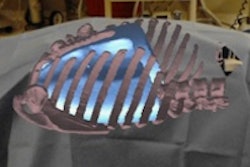
Virtual reality (VR) and augmented reality (AR) technology appear poised to have a major impact on diagnostic imaging interpretation, as well as procedure training, image-guided intervention, and interdisciplinary collaboration, according to a team from the University of Maryland School of Medicine. Learn how by clicking on our coverage of a recent webinar from the Society for Imaging Informatics in Medicine.
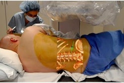
Researchers around the world are working to make that vision for VR and AR a clinical reality. For example, a team from the University of California, San Francisco is developing an imaging augmented reality software application that could be used to visualize planning for liver transplants and surgery. In addition, physicians at the University of Minnesota Masonic Children's Hospital have used VR models created with CT and MR images to plan the successful separation of conjoined twin babies. Get the details here.
We invite you to scroll through the links below for the rest of the news in advanced visualization, 3D printing, and CAD, all here in your Advanced Visualization Community.

