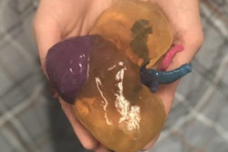
Researchers from China have created 3D-printed livers of pediatric patients with hepatic tumors and used these models to educate the parents -- improving their understanding of the condition and treatment options, according to a recent study published in the Journal of International Medical Research.
Using CT scans, the team of investigators from Guangzhou Medical University educated parents about the anatomy and physiology of the liver and hepatic tumors, as well as on the surgical procedure required for treatment and its associated risks. Providing an additional educational session with individually tailored 3D-printed livers improved the parents' scores on a questionnaire in every related category by more than 20% (J Int Med Res, February 13, 2018).
"The most remarkable advantage of a 3D-printed model is the straightforward exhibition of the liver organ, which is not possible on CT," senior author Dr. Yan Zou and colleagues wrote. "Participants significantly improved their understanding of all aspects of research issues [after education with 3D-printed models] compared with conventional methods."
Patient education
The capacity of 3D printing to provide tangible 3D visualization of internal organs has prompted its use in various fields of medical learning, not only to facilitate preoperative planning, training, and intraoperative implantation for surgeons but also to reinforce patients' understanding of their condition.
Clinicians most commonly use CT scans or MR images to supplement their explanation during patient education, especially in cases that require an invasive procedure, according to the authors. But patient-specific 3D-printed models could offer a novel tool that's easier for patients to understand than 2D medical images.
In this prospective study, Zou and colleagues oversaw the clinical consultations at Guangzhou Women and Children's Medical Center for seven children who had liver tumors and needed to undergo a partial hepatectomy. The first 20-minute meeting included the presentation of information using CT scans regarding the liver, hepatic tumors, and the details and risks of the patients' upcoming surgery.
Following this initial consult, the parents completed a questionnaire containing 22 true-or-false questions aimed at assessing their understanding of the liver in general, the child's medical condition, and the operation the child would undergo. Afterward, the same clinician who offered the information showed the parents a unique 3D-printed model of their child's liver, described the basic 3D printing process to them, and answered any questions about the process. To conclude the session, the parents answered the same questionnaire again.
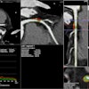
To make the 3D-printed livers, the researchers collected contrast-enhanced CT scans of the patients and processed and edited the scans -- including digitally segmenting vasculature -- using computer software (Mimics 14.01, Materialise). The software then automatically performed a surface extraction of the edited scans and converted them into the stereolithography (STL) format. Next, they transferred the files to a rapid prototyping STL printer (RS6000, Shanghai Union 3D Technology) at a commercial printing company for manufacturing and postprocessing.
Deeper understanding
All 14 parents achieved a higher score on the questionnaire after examining and learning about their child's 3D-printed liver. On average, the percentage of questions they answered correctly increased by 26.4% regarding basic liver anatomy, 23.6% for basic liver physiology, 21.4% for tumor features, 31.4% for the surgery, and 27.9% for risks associated with the surgery.
| Parent understanding after education with CT alone vs. CT plus 3D printing | |||||
| Topic | Median percentage of correct responses after presenting CT scans | Median percentage of correct responses after presenting CT scans and 3D-printed liver | |||
| Liver anatomy | 50% | 80% | |||
| Liver physiology | 50% | 70% | |||
| Tumor features | 60% | 80% | |||
| Partial hepatectomy | 60% | 90% | |||
| Risks associated with surgery | 60% | 90% | |||
The results suggest that showing 3D-printed livers comprehensively improved parents' education and understanding. One of the primary reasons for this improvement was that the 3D-printed models offered a more straightforward depiction of the liver than the CT scans, according to the authors.
What's more, neither the patients nor parents reported feeling uncomfortable upon seeing a physical representation of the liver, the authors noted. Rather, all participants gave positive feedback concerning the 3D-printed liver.
"The use of a 3D-printed liver model in this current study gave participants a much better idea of what would happen during surgery, which reassured participants, and the patients consequently felt more involved in the decision-making process," they wrote. "It would be interesting to see whether these positive responses had an impact on actual outcomes after surgery."
A marked limitation of the study was that the researchers presented the 3D-printed liver soon after the initial consultation. This afforded the parents the opportunity to hear relevant information again, which could have inherently improved their understanding and, thus, elevated their scores. In addition, fabricating the 3D-printed livers was time-consuming and expensive: It took roughly eight hours just to prepare the 3D model before sending it for printing at the external company, and the cost of each model was approximately $450.
"As 3D printing technology progresses and costs fall, patient-specific 3D printing may become standard for both clinical and educational purposes," they wrote. "Advances in technology and multipurpose use of models in treatment planning, trainee education, and patient education may also help improve cost-efficiency."


