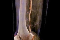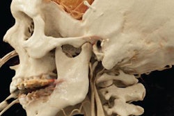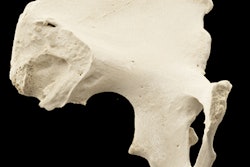
Cinematic rendering may help improve preoperative planning for complex surgeries in the stomach and kidneys by enhancing the surface detail of abdominal CT scans, according to research recently published online in Abdominal Radiology and the Journal of Computer Assisted Tomography.
Still relatively new to the field of radiology, cinematic rendering has been gaining traction as an intriguing alternative to conventional 2D interpretation of medical images. Its application has mostly been reserved for intricate cases involving highly vascularized anatomy and other difficult-to-visualize regions such as the aorta, due in part to the technique's novelty and high computing demands.
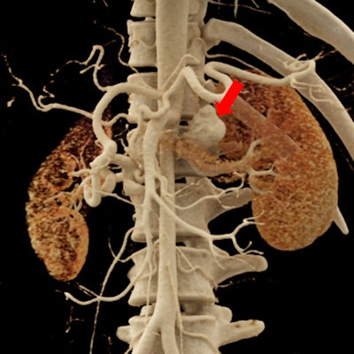
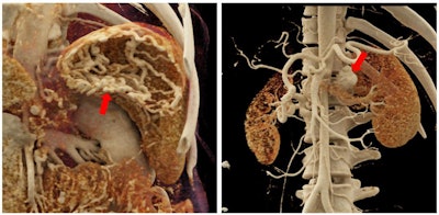 Cinematically rendered images of a stomach with extensive varices (left) and a relatively large saccular aneurysm near the left renal artery (right). Images courtesy of Dr. Elliot Fishman.
Some of the earliest users of cinematic rendering are 3D visualization expert Dr. Elliot Fishman and Dr. Steven Rowe, PhD, who are working with cinematic rendering software (syngo.via VB20, Siemens Healthineers) at Johns Hopkins University. In the current papers, they describe how they processed the abdominal CT scans of several patients with different types of renal and gastric pathologies, including kidney stones and infections, hydronephrosis, and various stomach neoplasms.
Evaluating cinematically rendered images of the kidney and stomach, they found that the photorealistic lighting and shadowing of these images allowed clinicians to appreciate the complex anatomy of the organs as well as subtle surface details, which they otherwise may have missed on standard CT scans (Abdom Radiol, May 19, 2018; J Comput Assist Tomogr, May 21, 2018).
In one case, the photorealism of cinematic rendering more clearly depicted the gastric mucosal pattern of an adenocarcinoma in a 61-year-old man's stomach than standard CT and 3D volume rendered images did. Cinematic rendering may thus aid "the detection of early, localized gastric adenocarcinomas, particularly those that are sessile or depressed from the gastric surface," the authors wrote.
"Cinematic rendering appears to recapitulate the advantages found in other 3D visualization techniques ... [including] volume rendering but with a more photorealistic appearance," they wrote. "Potential advantages of cinematic rendering in this context include improved preoperative planning via better understanding of the relative positions of anatomic objects within the imaged volume and facilitation of patient engagement and education as these images may be more intuitive for those without a formal medical background."
So far, case studies using the technique have yet to directly compare the benefits of cinematic rendering with those of other advanced visualization modalities for clinical evaluation and preoperative planning. The authors hope that continuing studies will explore the utility of cinematic rendering in a wide range of conditions to establish its potential diagnostic benefits.


