SAN FRANCISCO - Iterative reconstruction is a hot topic in CT imaging this year, lauded for its ability to reduce image noise by predicting and then replacing the missing data that plague noisy images. The image-reconstruction algorithms can deliver improved image quality along with dose reductions of one-third to one-half.
The use of one proprietary method in particular, adaptive statistical iterative reconstruction (ASIR) (GE Healthcare, Chalfont St. Giles, U.K.), has significantly reduced body imaging doses in several multicenter studies discussed at this week's International Symposium on Multidetector-Row CT.
In fact, ASIR has replaced filtered back projection entirely at all three Mayo Clinic sites in the U.S., where nearly 200 radiologists are using the technique in an ever-widening range of CT imaging applications.
Iterative evolution
Iterative reconstruction methods are quite different from the ubiquitous filtered back projection (FBP) technique that has traditionally been used for reducing image noise. In FBP, projection data are filtered and then pushed back along ray paths to form image pixels.
Iterative reconstruction is at once more straightforward and more complex, computing image data iteratively via statistical reconstruction algorithms and algebraic reconstruction techniques that predict the characteristics of missing image data using any number of factors, from nearby data in the same image to comparable anatomic images from a reference database.
The more complex iterative algorithms, known as model-based iterative reconstruction (MBIR), improve image quality significantly but are computationally dense -- and are therefore impractical for daily clinical use, pending the development of faster processors in the next several years.
Designed as a kind of compromise technique that can be processed quickly, GE's ASIR method cuts computing time by eliminating system optics modeling in the iterative algorithm, concentrating on statistical analysis instead.
In early studies, the use of ASIR has been found to enable dose reductions of one-third to one-half while improving image quality. Other manufacturers are said to be working on similar methods.
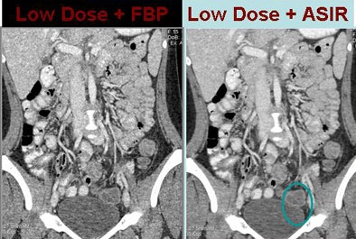 |
| In a15-year-old patient presenting to the emergency department to rule out appendicitis, low-dose scan with FBP reconstruction was noisier than follow-up imaging using the same dose with ASIR reconstruction. All images and data courtesy of Dr. Amy Hara. |
Mayo experience
"We've been using ASIR for the last year and found it helpful in any image where you want to decrease noise," said Dr. Amy Hara, radiologist at the Mayo Clinic in Scottsdale, AZ, in a Wednesday presentation at the symposium. Applied primarily to low-dose imaging, "it's also helpful for obese patients, whether or not you're able to use a low-dose technique, and ... for pancreatic imaging as well," she said.
The use of ASIR reconstruction, which can be scaled to the desired strength from 10% to 100%, can cut image noise by as much as 65% while improving image quality, edge, and lesion conspicuity.
"When we do quantitative measurements, the ASIR images have quantitatively less noise as well," Hara said. Potential pitfalls include liver edge artifact, a plastic or artificial appearance of the CT images if dialed up too high, and excessive automatic dose reduction in thin patients.
Implementing the technique is simple. It replaces FBP in the workflow and requires about the same processing time, Hara said. For a given application, the radiologist simply increases the image noise index; for example, from 21 to 30. This causes the automatic exposure control to reduce the mAs to match the new noise level. Then, when reconstructing the images, ASIR is applied in lieu of FBP.
"For most, I think the real question is how does a low-dose examination with ASIR compare to routine dose on CT with filtered back projection when we're talking about using this clinically for all of our patients," she said. "We had a lot of concern about either missing lesions or misdiagnosing lesions."
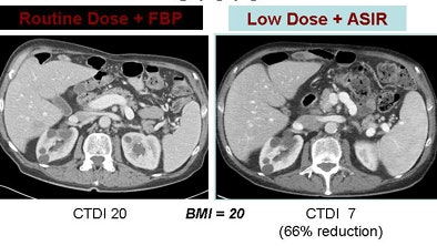 |
| A rectal cancer patient underwent abdominal CT with a normal-dose scan plus FBP (left) and follow-up with a low-dose technique using ASIR reconstruction (right). Despite a dose reduction of 66%, renal cysts are easily visualized in the low-dose technique. |
In the year they've been working with the technique, Dr. Alvin Silva, Hara, and colleagues have performed several studies on patients undergoing abdominal CT exams, including research submitted for publication.
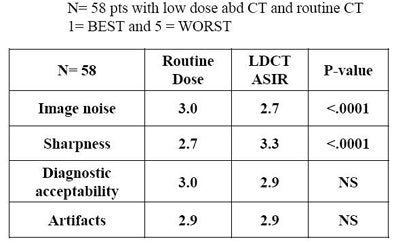
One study examined 58 patients who underwent a routine-dose abdominal CT exam with FBP reconstruction and later were followed-up with low-dose CT plus ASIR.
A subjective image quality evaluation by two abdominal radiologists found that the low-dose ASIR images had comparable image quality, with slightly less image noise than the normal-dose images and slightly poorer liver-edge sharpness scores, Hara said.
 |
| Silva, Hara, and colleagues examined subjective image quality in 58 patients who underwent abdominal CT with a normal dose and FBP. The patients later received a follow-up scan using approximately one-third less dose and ASIR reconstruction. Noise and diagnostic acceptability results were comparable. |
 |
Getting used to ASIR
"I think the biggest hurdle for radiologists looking at ASIR-reconstructed images is that the images look different," Hara said. "You can use anywhere from 10% to 100% ASIR, and if you dial up all the way to 100%, you start to get kind of a plastic, artificial look to the CT images because there's so much less image noise."
Whether 100% ASIR are lacking diagnostically has not really been determined, but radiologists' initial reaction can be that the images are not what CT is supposed to look like, Hara said. Many users consider 50% ASIR a good balance between reduced noise and a natural appearance, while others prefer 30% or 40%.
"It's very subjective," she said. But "having done ASIR reconstructions for a year, you get used to a new normal, and if you look back to the filtered back projections you really notice the image noise."
Another study looked at 36 patients who underwent a single low-dose abdominal CT exam, either using FBP or ASIR reconstruction. The images were examined by two abdominal radiologists blinded to the reconstruction technique, who graded the images subjectively on a scale of 1 (best) to 5 (worst). The ASIR images were significantly better in subjective image noise, subjective image quality, and ROI noise measurements, Hara said.
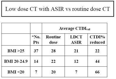
Hara and colleagues have also found that image quality scores were lower in thin patients, because increasing the image noise index resulted in excessive dose reduction of as much as 75%. This problem was corrected by increasing the minimum mAs on the scanner from 50 to 100, enabling more consistent dose reduction percentages across a range of patient body sizes.
 |
| Silva, Hara, and colleagues examined 57 patients who underwent routine-dose abdominal CT with filtered back projection (RD FBP) and low-dose CT with ASIR reconstruction (LD ASIR). Two abdominal radiologists blinded to the reconstruction method graded image quality on a scale of one to five. LD ASIR was superior to RD FBP in qualitative and quantitative image noise, the methods were equivalent in diagnostic acceptability and the presence of beam-hardening artifact, and RD FBP was superior in liver edge sharpness. The overall dose reduction was 34%. |
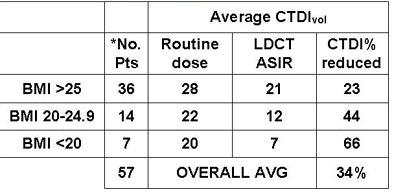 |
"In the last year for routine body CT we've achieved dose reductions of anywhere from 23% to 66% depending on size, averaging about 30%," Hara said. "In our preliminary studies, it doesn't look like [ASIR] reduces lesion conspicuity or detection, which was a big issue that we wanted to solve, and we're currently using anywhere from 30% to 40% ASIR for body CT."
By Eric Barnes
AuntMinnie.com staff writer
May 20, 2009
Related Reading
New algorithm sharpens coronary artery images, July 14, 2008
Strategies for reducing 'dose creep' in digital x-ray, April 11, 2007
CARS report: Fuzzy theory yields sharper CTA images, June 27, 2008
3D development outpaces facilities' readiness, June 23, 2008
Video game technology speeds beating-heart surgery, June 9, 2008
Copyright © 2009 AuntMinnie.com


