Color Doppler ultrasound can be reliably used to diagnose carpal tunnel syndrome (CTS), while an image processing method utilizing power Doppler pixel information can also quantitatively evaluate its severity, according to research published online in Radiology.
A prospective study from Tehran University of Medical Sciences in Iran found that color Doppler ultrasound could provide sensitivity and specificity similar to electrodiagnostic testing (EDT). In addition, an image processing program developed by researchers at the university was able to achieve good concordance with EDT for evaluating carpal tunnel severity.
"Color Doppler US can be used as a noninvasive alternative to EDT in both the diagnosis of CTS (by determining the presence of intraneural vascularity) and the assessment of its severity (by determining the pixel numbers in the intraneural vascular area with use of an image processing program); Color Doppler imaging may be used in parallel to other imaging modalities (e.g., grayscale US), which have already shown promising results," the authors wrote.
The diagnosis of carpal tunnel is routinely confirmed with EDT technologies such as electromyography and nerve conduction studies. Because EDT suffers from concerns over diagnostic accuracy, imaging modalities are increasingly being evaluated for their potential usefulness in this application, according to the researchers.
In carpal tunnel syndrome, the intraneural microvasculature of the median nerve is prominent and dilated due to inflammation and ischemia within the median nerve. As a result, the researchers hypothesized that color Doppler ultrasound evaluation of intraneural vasculature might aid in detecting CTS, according to co-author Dr. Omid Khalilzadeh.
In a pilot evaluation of patients with carpal tunnel, the researchers observed that most had a detectable intraneural vascularity, and the degree of vascularity positively correlated with the severity of the patients' symptoms. They also found that intraneural vascularity was often not evident in healthy individuals.
To provide quantitative information, the researchers believed they could estimate the quantity of intraneural blood vascularity by determining the number of pixels in areas of blood flow on recorded power Doppler ultrasound images. After designing an image processing program to perform that task, they evaluated the relationship of those pixel numbers and the severity of carpal tunnel syndrome in a study of 101 patients with definite clinical evidence of CTS and 55 healthy volunteers from September 2009 to March 2001 (Radiology, September 7, 2011).
An experienced neurologist determined if there was clinical evidence of carpal tunnel, and the diagnosis was confirmed by an experienced hand surgeon. The researchers excluded any participants with a history of surgical treatment for CTS, wrist trauma or fracture, or other conditions associated with neuropathy. Patients with clinical findings of any other neuropathies were also excluded.
Healthy volunteers were chosen from hospital staff. The study team did not find a significant difference between patients and control subjects with regard to sex ratio or age distribution.
EDT (standardized electromyography and nerve conduction studies) were performed in all participants using a 10-channel system (Oxford Instruments Medical). Within one week of EDT, all ultrasound exams were performed by one radiologist with approximately five years of musculoskeletal imaging experience.
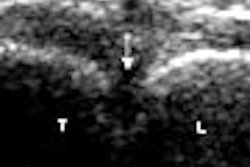
Blinded to whether the subject was in the control or patient group, the radiologist performed color Doppler ultrasound on a Sonoline Antares scanner (Siemens Healthcare) using a 9- to 13-MHz linear-array probe, and a setting suitable for low-flow vessel detection. Power Doppler ultrasound followed the conventional color Doppler study.
An image processing program was then applied to the power Doppler images, which were recorded directly in JPEG format by the ultrasound system. Developed by the research center's medical engineering team, the program provides quantitative assessment of carpal tunnel syndrome severity by counting the number of pixels in parts of images colored red, which represent the intraneural vascular area.
Performance in carpal tunnel syndrome
|
The difference was not statistically significant (p > 0.05).
In other findings, intraneural vascularity was seen in all patients with moderate (26) or severe (21) CTS and in 32 (91.4%) of those with mild carpal tunnel syndrome.
As for assessment of CTS severity, the median sum of pixels in the intraneural vascular area was 281 (range, 139-471) in the control group, compared with 379 (range, 105-2201) in the patient group. The difference was statistically significant (p < 0.001).
Khalilzadeh said that other institutions and vendors can access their image processing method and Matlab software program codes for free.
The next phase of their research will involve applying this method for other compression neuropathies, he told AuntMinnie.com.
"We suggest that this method can be easily adopted and used for evaluation of all compression neuropathies in body," he said.
The researchers will also evaluate the benefits of their noninvasive method in following up carpal tunnel patients after medical and surgical treatments.


