Dear Advanced Visualization Insider,
3D printing has traditionally focused on bone imaging, with other applications largely relegated to research. But doctors and engineers in Illinois have broken the mold to bring this exciting technology into clinical use in one of the most challenging surgical jobs in medicine: repairing complex congenital heart defects in young children.
The team has developed a technique that creates whole-heart 3D models from high-resolution MR images and a new generation of 3D printers and software. Surgeons working on the project believe the models are the only way they can learn what they'll really be facing in surgery before young patients get on the table. Learn more in this issue's Insider Exclusive, based on a presentation given today in Chicago at the American Heart Association meeting and brought to you before our other members can access it.
Is it time for a revolution? Finding and analyzing specific locations in 3D images is slow and tedious under the best of circumstances. But developers of a new software platform known as Virtual Finger say their new algorithm can navigate reliably and almost instantaneously with the click of a mouse. Virtual Finger knows the precise depth and location of your mouse click, and the whole thing runs on a laptop. What do the algorithm and human vision have in common? Click here to find out.
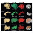
Accurate segmentation of brain tissue in MRI -- to distinguish gray from white matter and cerebrospinal fluid and such -- is the first thing neurologists need to get a handle on almost any brain pathology, from multiple sclerosis to Parkinson's and Alzheimer's diseases. However, automated segmentation has been a tough nut to crack, owing to some specific MRI artifacts and, even more substantially, to extensive signal overlap between tissue types. Researchers from Texas think they've solved all that with a new algorithm that works far better than previous methods. It's not only accurate, it's computationally efficient, they say. Learn more here.
Can black and white and the colors of the rainbow find true happiness together on the same medical image display? They can, according to a new generation of displays that unites previously segregated screen types as well as any Benetton ad -- and hopefully erases some painful ergonomic strains in the process. You'll find the rest of the story here.
We invite you to scroll through the links below for more news about the ways computer visualization is making radiology easier and more accurate. And stay tuned for the latest news on the imaging revolutions happening every day, right here in your Advanced Visualization Community.


