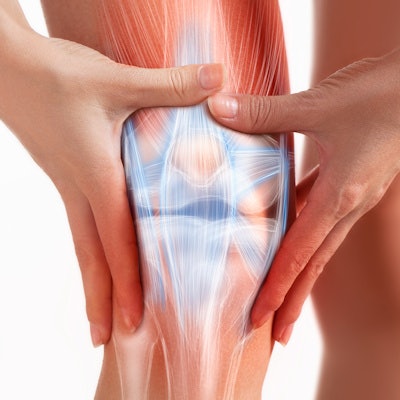
The RSNA has published a new Quantitative Imaging Biomarkers Alliance (QIBA) profile that aims to facilitate more reliable and reproducible MRI-based measurements of cartilage degeneration in the knee.
In an article posted online on September 7 in Radiology, the RSNA's Musculoskeletal Biomarker Committee shared the new QIBA profile for standardized MRI-based knee cartilage compositional imaging that includes two advanced MRI techniques: spin-lattice relaxation time constant in rotating frame (T1ρ) and T2 mapping.
The committee concluded that T1ρ and T2 are measurable with 3-tesla MRI with a within-subject coefficient of variation of 4%-5%. What's more, the group found that a 14% or more measurable increase in T1ρ and T2 indicates a minimum detectable change that can be used for defining response and progression criteria for quantitative cartilage imaging, according to the authors led by Dr. Majid Chalian of the University of Washington.
However, if only an increase in T1ρ and T2 is expected due to progressive cartilage degeneration, then a 12% increase would be considered to be the minimum detectable change.
Although MRI-based cartilage compositional analysis is a promising approach for revealing biochemical and microstructural changes in the early stages of osteoarthritis, clinical applications have been limited so far. T1ρ and T2 mapping have been established for assessing cartilage composition, but only T2 mapping is currently available commercially, according to the researchers.
As a result, the committee developed a QIBA profile for MRI-based knee cartilage compositional imaging by reviewing published studies in the literature. The researchers also hope that the profile will facilitate clinical applications by promoting a shift from manual segmentation of MRI sequences to an automated approach. Several machine-learning applications for automatic cartilage segmentation are promising, according to Chalian.
"We are hoping that a machine learning approach can segment out the cartilage, and then we can apply this profile on the segmented cartilage so that we can make things go fast in busy clinical settings," said Chalian in a statement from the RSNA.
In an accompanying editorial, Dr. Richard Kijowski of the New York University Grossman School of Medicine noted that the new QIBA profile is an important first step toward standardizing compositional cartilage MRI biomarkers.


