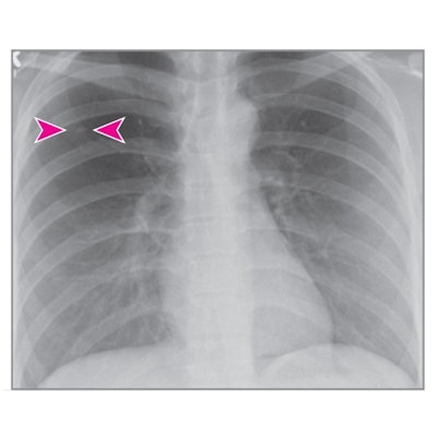
An artificial intelligence (AI) algorithm can improve the diagnostic performance of radiologists for detecting pulmonary nodules on chest x-rays, according to a study published December 29 in JAMA Network Open.
An international group that included scientists from Siemens Healthineers tested the company's AI-Rad Companion Chest X-ray algorithm to see if it improved accuracy among junior and senior radiologists. The team found that AI-aided interpretation of chest radiographs resulted in a lower number of missed and false-positive pulmonary nodules among a group of nine participating radiologists.
"AI-aided interpretation was associated with significantly improved detection of pulmonary nodules on chest x-rays as compared with unaided interpretation of chest x-rays," wrote corresponding author Dr. Mannudeep Kalra, an associate professor of radiology at Harvard Medical School in Cambridge, MA.
The detection of lung cancer nodules on chest radiographs has been a focus of AI research for 20 years. Several recent studies suggest AI can improve the performance of radiologists for identifying pulmonary nodules on chest x-rays, yet previous studies have been limited in scope due to the lack of multisite readers with different levels of clinical experience, detection difficulties, and settings, according to the authors.
In light of these limitations, the group designed a study that included 100 posterior-anterior images acquired on x-ray units from nine different vendors, including Carestream Health, Fujifilm Medical Systems, GE Healthcare, and Varian Medical Systems. The images were from two different sources -- an ambulatory healthcare center in Germany and a lung image database in the U.S. -- acquired between 2000 and 2010.
Images were selected to represent nodules with different levels of detection difficulties (from easy to difficult). Two thoracic radiologists established the ground truth and nine junior and senior radiologist readers from Germany and the U.S. independently reviewed all images in two sessions, unaided and AI-aided.
The analysis included metrics for sensitivity, specificity, accuracy, and receiver operating characteristics curve area under the curve (AUC).
Results showed the mean detection accuracy across all nine radiologists improved by 6.4% with AI-aided interpretation compared with unaided interpretation. Partial AUCs within the effective interval range of a 0 to 0.2 false-positive rate improved by 5.6% with AI-aided interpretation.
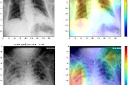
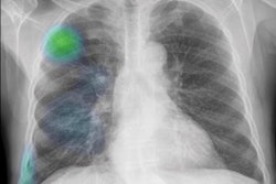
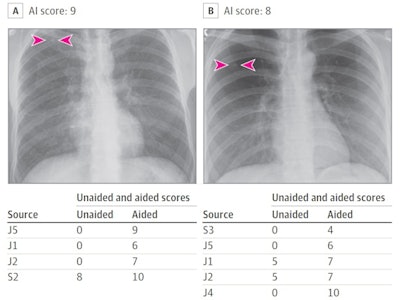 (Panel A) Artificial intelligence (AI) helped detect right upper lobe nodules (arrowheads) for three junior (J) radiologists, as well as improved the confidence score for one senior (S) radiologist (score > 5 implies positive finding). Likewise, AI-aided interpretation led to detection of missed right upper lobe nodules (arrowheads) for the radiograph in panel B for two junior radiologists and improved confidence of two junior radiologists. Image courtesy of JAMA Open Network.
In addition, junior radiologists saw greater improvement in sensitivity for nodule detection with AI-aided interpretation as compared with their senior counterparts (12% vs. 9%), while senior radiologists experienced similar improvement in specificity (4%) as compared with junior radiologists (4%).
"[AI-Rad Companion Chest X-ray] was associated with improved detection of pulmonary nodules on chest radiographs compared with unaided interpretation for different levels of detection difficulty and for readers with different experience," the authors wrote.
The study did not assess the clinical relevance of pulmonary nodules with AI-aided interpretation as opposed to unaided interpretation, and despite their inferiority to low-dose CT images, chest x-rays are frequently used for early detection of pulmonary nodules, the researchers noted.
Pulmonary nodules can be easily missed when they are subtle, small, or in difficult areas, they added.
"Given the high miss rate and importance of identifying pulmonary nodules on chest radiographs, AI algorithms such as the one assessed in our study and those tested in prior studies can play an important role in improving quality and diagnostic value of radiographs in patients with pulmonary nodules," the group concluded.
The AI-Rad Companion Chest X-ray software is pending regulatory clearance. Siemens received U.S. Food and Drug Administration (FDA) clearance in September 2019 for AI-Rad Companion Chest CT as an aid for imaging of the lungs, heart, and aorta.


