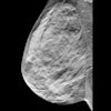
AI-supported mammography finds more breast cancers during screening and reduces interval cancer diagnosis by 12% in the years following screening, suggest findings published January 29 in The Lancet.
A team led by Jessie Gommers, PhD, from Radboud University Medical Center in Nijmegen, the Netherlands, found that AI-supported mammography screening showed consistently favorable outcomes compared with double reading. These include finding a comparable number of interval cancers and having higher sensitivity.
“These findings suggest that with the support from AI breast imagers can safely and efficiently increase the sensitivity in screening,” co-author Kristina Lång, MD, PhD, from Lund University and Skåne University Hospital in Sweden, told AuntMinnie.
While prior research suggests that AI can improve breast cancer screening via improved disease detection and workload reduction, its effect on interval cancers is unknown.
The Mammography Screening with Artificial Intelligence (MASAI) trial is currently the only randomized controlled trial evaluating AI-supported mammography screening to have reported results and is the first such study to be completed. Published results showed a 44% reduction in screen-reading workload for radiologists and a 29% increase in cancer detection without increasing false positives.
Gommers, Lång, and colleagues compared the interval cancer rate in AI-supported mammography screening with standard double reading with no AI help in a randomized, controlled, noninferiority, single-blinded, population-based screening accuracy trial.
The team randomly assigned 105,915 women to either the AI assistance or standard double-reading group in a 1:1 ratio. AI helped in triaging exams to single or double reading by radiologists and for detection support.
AI-assisted mammography led to noninferior interval cancer rates versus double reading, as well as superior sensitivity and comparable specificity.
Comparison between AI-assisted mammography, standard double reading | |||
Measure | Double reading | AI-assisted mammography | p-value |
Interval cancer rate (per 1,000 women) | 1.76 | 1.55 | 0.41 |
Invasive interval cancers (per 1,000 women) | 89 | 75 | N/A |
T2+ interval cancers (per 1,000 women) | 48 | 38 | N/A |
Nonluminal A interval cancers (per 1,000 women) | 59 | 43 | N/A |
Sensitivity | 73.8% | 80.5% | 0.03 |
Specificity | 98.5% | 98.5% | 0.88 |
The team noted that the interval cancer rate measure achieved a noninferior proportion ratio of 0.88.
It also reported that AI-assisted mammography’s sensitivity was consistent across age, breast density, and for invasive cancer. However, AI aid did not lead to improved sensitivity for in-situ cancer (90.7% vs. 91.8% for double reading; proportion ratio, 0.99).
Lång said the findings suggest a shift toward earlier detection of clinically relevant cancers, reflected in fewer aggressive or advanced interval cancers.
“In addition, the use of AI reduced the screen-reading workload for radiologists by almost half compared with standard double reading,” she said. “This reduction is particularly relevant in the context of a growing shortage of breast radiologists.”
Lång also told AuntMinnie the team is currently studying how AI influences the performance of breast radiologists during screen-reading.
“By running the same AI algorithm on the control group examinations, we can compare performance in low- and high-AI risk examinations and test whether AI reduces false positives in low-risk examinations and reduces false negatives in high-risk examinations,” she said. “We are also conducting a detailed review of interval cancers to determine which cancers were missed and whether they were visible in retrospect.”
“Additionally, we plan to study outcomes in the next screening round to see how AI may affect subsequent screenings and the longer-term effect on the incidence of advanced cancers,” Lång noted. “We are also performing cost-effectiveness analyses to understand how AI could impact healthcare costs.”
Read the full study here.