An AI algorithm significantly improves radiologist performance for identifying early lung cancer on low-dose chest CT -- with benefits across nodule types, screening contexts, and experience levels, according to a study published January 29 in the Journal of the American College of Radiology.
The findings "support integrating AI into chest CT workflows to enhance early-stage lung cancer detection," noted a team led by Jamie Schroeder, MD, DPhil, of Georgetown University Medical Center in Washington, DC.
"[We found that] AI assistance increased radiologist sensitivity by 24.3% without meaningfully raising false positives," the group wrote.
Schroeder and colleagues investigated whether a deep learning-based AI system would improve radiologists' performance in detecting pulmonary nodules on CT. The team trained an AI algorithm with a dataset enriched with early-stage lung cancers and compared its standalone sensitivity and specificity against 16 radiologist readers.
Each reader interpreted 340 CT scans with and without AI, separated by a one-month break. The dataset included 209 screening and 131 nonscreening cases; of these, 133 showed lung cancer, 61 showed benign noncalcified nodules equal to or less than 4 mm, and 146 were normal. The team assessed radiologist performance using the localization receiver operating characteristic (LROC) area under the receiver operating curve (AUC) measure (LROC AUC).
The group reported the following:
Comparison of radiologist identification of early lung cancer, with and without AI assistance measured by LROC AUC | ||
Measure | Without AI assistance | With AI assistance |
| Cancer detection, overall | 0.65 | 0.76 |
| Cancer detection across all nodule types | 0.73 | 0.83 |
| Sensitivity | 0.59 | 0.73 |
| Specificity | 0.92 | 0.91 |
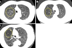
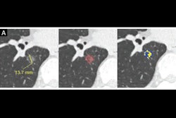
Mean interpretation time decreased from 133 seconds to 115.9 seconds, a reduction of 12.9%, according to the researchers. They found the greatest performance gains in screening settings, particularly for small nodules and cancers under 10 mm, and AI alerts identified lung lesions that "should have triggered a positive National Lung Screening Trial (NLST) screen but were missed during initial radiologist assessments."
"The AI system significantly improved radiologists' sensitivity and efficiency in detecting lung nodules, including clinically challenging early-stage cancers, supporting its integration into chest CT workflows to enhance screening performance," they concluded.
Access the full article here.