J Nucl Med 2002 May;43(5):632-4
Comparison of (123)I and (131)I for whole-body imaging in thyroid cancer.
Sarkar SD, Kalapparambath TP, Palestro CJ.
We compared the diagnostic sensitivities of (123)I and (131)I whole-body imaging
in differentiated thyroid cancer. METHODS: Twelve thyroidectomized patients (3
previously treated with (131)I) were studied. After a period of thyroid hormone
withdrawal, whole-body imaging was performed approximately 24 and 72-96 h after
administration of 74-185 MBq (2-5 mCi) (123)I and 111-185 MBq (3-5 mCi) (131)I,
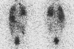
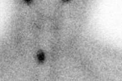
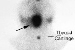
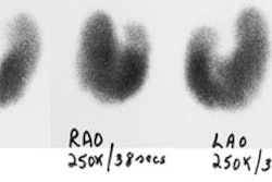
respectively. RESULTS: Both (123)I and (131)I revealed residual thyroid tissue,
present in 9 patients. (131)I detected metastases in 5 studies of 4 patients. In
4 of 5 studies, (123)I missed metastases shown by (131)I in 8 body regions
including the neck, mediastinum, lungs, and bone and detected 3 other sites of
metastasis only in retrospect. No lesion was better seen with (123)I than with
(131)I. CONCLUSION: Although (123)I is adequate for imaging residual thyroid
tissue, it appears to be less sensitive than (131)I for imaging thyroid cancer
metastases.