D-Transposition of the Great Vessels
Case submitted by Dr. Scott D. Flamm)
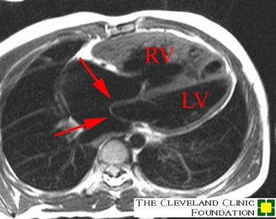
This first image below is a four chamber spin echo image showing the markedly hypertrophied and dilated right ventricle (anterior ventricular chamber) and the thin walled, smaller left ventricle (posterior ventricular chamber). The red arrow points to the "Mustard" inter-atrial baffle that redirects blood flow within the atria. Pulmonary venous return is directed towards the tricuspid valve and then into the right ventricle (and subsequently the aorta), while the systemic venous return from the SVC and ICV is directed towards the mitral valve, into the left ventricle (and susequently into the pulmonary arteries).

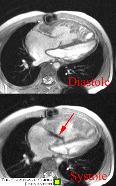
The second image is a cine-GRE. The red arrow demonstrates the systolic tricuspid regurge jet that courses along the posterior aspect of the leaflet. D-TGA's often get moderate to severe tricuspid regurge because the tricuspid annulus dilates secondary to right ventricular dilatation (the RV is not meant to severe as a "systemic" ventricle)


