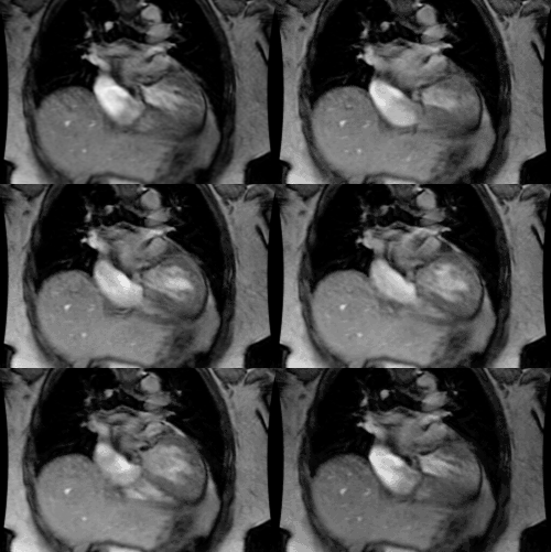
Radiographics 1997 Jan;17(1):145-153
Malignant tumors of the heart and great vessels: MR imaging
appearance.
Mader MT, Poulton TB, White RD
Department of Diagnostic Radiology, Aultman Hospital, Canton,
OH 44710, USA.
Fortunately, primary tumors of the heart and great vessels are rare. These primary tumors include angiosarcoma, malignant
fibrous histiocytoma, high-grade and pleomorphic sarcoma, and paraganglioma with pericardial and myocardial invasion.
Symptoms are often nonspecific and include chest pain and dyspnea. Although these tumors are often diagnosed with
echocardiography and computed tomography, magnetic resonance (MR) imaging currently appears to be the imaging
modality of choice because of its diverse capabilities, which include multiplanar imaging for excellent anatomic definition of the
heart, pericardium, mediastinum, and lungs; improved morphologic differentiation between tumor tissue and surrounding
cardiovascular, mediastinal, or pulmonary tissues; dynamic imaging with a gated cine-loop acquisition; and assessment of
tissue perfusion. The use of gadopentetate dimeglumine is helpful in achieving tumor enhancement on MR images but is not
required. MR imaging is also useful in assessing tumor response
to surgery, radiation therapy, and chemotherapy.
MeSH Terms:
Adult
Aortic Diseases/diagnosis*
Female
Heart Neoplasms/diagnosis*
Hemangiosarcoma/diagnosis
Histiocytoma, Fibrous/diagnosis
Human
Magnetic Resonance Imaging
Male
Middle Age
Paraganglioma/diagnosis
Pulmonary Artery*
Sarcoma/diagnosis
Vascular Neoplasms/diagnosis*
PMID: 9017805, MUID: 97170276


