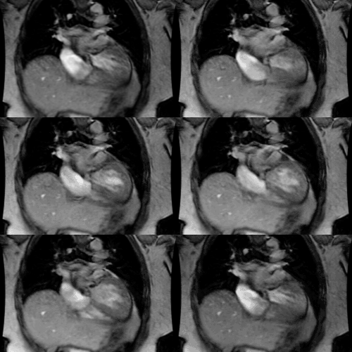
Ebstein's Anomaly:
View cases of Ebstein's anomaly
Clinical:
Ebstein's anomaly accounts for only 0.5 to 0.7% of cases of congenital heart disease [3]. It results from inferior displacement of the tricuspid valve (*the septal and posterior leaflets are usually affected*) below its normal annulus into the right ventricle resulting in outflow obstruction and decreased functional right ventricular volume. While the anterior leaflet is usually attached in its normal position, it is often large and dysplastic [2]. The portion of the RV proximal to the abnormal valve is incorporated into the RA (an atrialized Rright ventricle). This creates a functional obstruction to RA emptying with increased RA pressures and RA to LA shunting via a patent foramen ovale or ASD). Affected patients have associated tricuspid insufficiency (regurge). The amount of cyanosis and age at presentation are related to the amount of pulmonary blood flow. About 10% of cases are associated with chronic maternal lithium use. Associated cardiac lesions include a right to left shunt (PDA/ASD) and conduction disturbances- most commonly Wolff-Parkinson-White syndrome and right bundle branch block [2].
The clinical presentation is variable [2]. If left untreated, about 50% of the affected patients die in the first year of life, usually as a result of CHF. However, many affected adult patients are asymptomatic [2].
X-ray:
On CXR the heart can be very enlarged and develops a 'box shape' or "balloon shape" due to marked RAE and a narrow vascular pedicle (Ebstein's anomaly is the only cyanotic congenital malformation of the heart in which both the aorta and the pulmonary trunk are smaller than normal [3]). The pulmonary vascularity is decreased (but it can be normal and these patients will be acyanotic) and the pulmonary trunk is hypoplastic (concave pulmonary artery segment).
Leaflet displacement can be assessed on MR imaging [4]. Leaflet
displacement is determined by measuring the distance of the septal
leaflet to the anterior mitral valve leaflet [4]. This distance
should be greater than 15 mm in children younger than 14 years or
greater than 20 mm in adults [4]. When this distance is indexed to
body surface area, a value greater than 0.8 mm/cm2 is diagnostic
of Ebstein anomaly [4].
REFERENCES:
(1) Pediatric Clinics of North America 1999; Waldman JD, Wernly
JA. Cyanotic congenital
heart disease with decreased pulmonary blood flow in children.
46(2): 385-402
(2) Radiology 2004; Beerepoot JP, Woodard PK. Case 71: Ebstein anomaly. 231: 747-751
(3) Radiographics 2007; Ferguson EC, et al. Classic imaging signs
of congenital cardiovascular abnormalities. 27: 1323-1334
(4) Radiographics 2015; Malik SB, et al. The right atrium: gateway to the heart- anatomic and pathologic imaging findings. 35: 14-31


