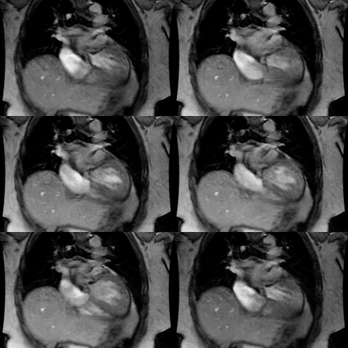
Atrial Fibrillation:
Arrhythmogenic foci originating within the pulmonary veins are important causes of both paroxysmal and persistent atrial fibrillation [1]. In most individuals, sleeves of left atrial myocardium extend into the pulmonary veins for a distance of 2-17 mm [1]. These sleeves are longest in the superior pulmonary veins and thickest at the venoatrial junction of the left superior pulmonary vein [1]. Hence, almost 50% of the foci arise in the left superior pulmonary vein [1]. Transvenous radiofrequency ablation of these foci is performed to electrically disconnect them from the left atrium [1].
MDC can be performed to evaluate for pulmonary venous anomalies and to define ostial orientation and distance from each ostium to the bifurcation of each pulmonary vein [1].
REFERENCES:
(1) AJR 2004; Cronin P, et al. MDCT of the left atrium and pulmonary veins in planning radiofrequency ablation for atrial fibrillation: a how-to guide. 183: 767-778


