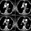
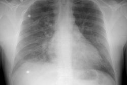
Single detector exam:
A bolus contrast enhanced exam should be performed from the level of the aortic arch to approximately 2 cm below the level of the inferior pulmonary veins with the patient scanned at end inspiration [37]. Hyperventilation before the start of the exam (consisting of a few deep breaths) is recommended as it will facilitate prolonged breath holding. Patients unable to maintain a breath hold can be scanned while gently breathing to reduce respiratory motion artifacts.
The scan is performed from just above the lower hemidiaphragm to the aortic arch (caudocranial direction) during an infusion of 150-200 ml of 30% contrast material (3-5 ml/sec) with a 15-20 sec scan delay (some centers use 60% iodinated contrast and a lower injection rate). Alternatively, a small test injection (15-20 cc) can be used to determine optimal timing of the scan- this is particularly beneficial in patients with known or suspected cardiac dysfunction [90]. The time to peak enhancement plus 5 seconds is used as the time delay for the diagnostic scan. The extra 5 seconds allows for opacification of the distal small arteries and provides a margin for error. For patients with right ventricular failure or pulmonary hypertension a longer scan delay is required (15-18 seconds) [37]. Scanning caudo to cranial and using a lower injection rate reduces streak artifacts from concentrated contrast material in the superior vena cava which may obscure the adjacent right main pulmonary artery. Also, caudo-cranial imaging will minimize motion artifacts due to respiration that tend to be greatest in the lung bases and less significant in the upper lungs.
A 3 mm collimation, table feed of 5 mm/sec (pitch of 1.7), 240-300 mA, with a 20 to 30 second exposure, and 1.5 mm overlapping reconstruction images read at the workstation will provide excellent image quality [16]. Patients not able to hold their breath for 20 to 30 seconds can be imaged with shorter scan times (10 mm/sec table feed with 5 mm collimation and a 10 second breath hold [pitch of 2]), but there is a decrease in spatial resolution [17]. Patients on a respirator can be imaged during a forced period of apnea or while breathing at a minimal tidal volume and respiratory rate [18].
For sub-second scanners, improved evaluation of subsegmental arteries can be obtained with the use of 2 mm collimation, 0.75 sec per revolution, and a pitch of 2 (ie: table speed of 4 mm per sec) [8,37]. Overlapping reconstruction images are not required when using this narrow collimation [37]. Electron beam CT has minor advantages in analyzing paracardiac arteries due to a reduction in motion artifact and higher contrast enhancement, but the images suffer from slightly increased noise [42]. Images should be reconstructed using a 180 degree linear interpolation algorithm to decrease partial volume effects [37].


