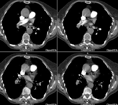
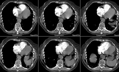
Case 8: Central PE with right heart strain.
The images below are from an elderly patient with a large embolic clot burden. There is a right main pulmonary artery embolism and the right descending pulmonary artery is completely obstructed (first image). The patient had multiple other emboli bilaterally. Note the right ventricular dilatation (in which the greatest short axis measurement of the right ventricular cavity is wider than the left ventricular cavity and the deviation of the interventricular septum toward the left ventricle (second image)- these findings are indicative of right heart strain and an increased mortality risk.


