Dear Orthopedic Imaging Insider,
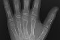
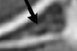
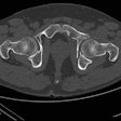
Radiography has been the initial modality used for assessing fractures in the long bones, but conventional x-ray can have a tough time differentiating stress fractures -- those caused by normal use -- from pathologic fractures, which are the result of bone tumors.
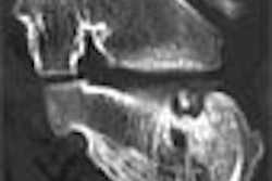
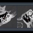
Cross-sectional imaging modalities like CT and MRI are frequently used after x-ray to provide additional diagnostic information, but few studies have been done that detail the advantages and drawbacks of each. In this edition of the Orthopedic Imaging Radiology Insider, we detail research from Johns Hopkins University that tries to do just that.
A group from Johns Hopkins found that in general, MRI performed with greater accuracy than CT or radiography in differentiating stress fractures from pathologic fractures. But CT still had information to contribute in several areas of fracture assessment, and radiography should still be used as a front-line imaging modality, they said. Read all about it in our Insider Exclusive, which you're receiving before other AuntMinnie members. Click here to get to the story.
In other recent articles in the Orthopedic Imaging Digital Community, a study by researchers at the University of Pennsylvania indicates that sickle cell disease significantly reduces whole-body bone mineral content in children.
In another study, Danish researchers found that adding the antiarthritis drug anakinra to methotrexate (MTX) does not prevent joint erosion as measured by MRI in patients with severe rheumatoid arthritis (RA).
Click on the links below for more orthopedic imaging news. If you have any ideas or suggestions for stories, please feel free to e-mail me at [email protected].
