Dear Orthopedic Imaging Insider,
In this issue, we feature two articles on using MRI as the proverbial hall monitor in the musculoskeletal system. Our Insider Exclusive is a report from the 2006 RSNA meeting on MR assessment of Achilles tendon repair. In their talk, researchers from Munich, Germany, laid out their imaging protocol, as well as what to look for on MR exams performed after treatment. To learn more, click here.
In a second story, physicians in California report that MR has the edge over ultrasound and x-ray for tracking bone erosion from rheumatoid arthritis. Click here for the details.
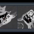
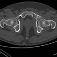
Then check out an article on Chance-type spinal fractures and how to make sense of the spectrum of imaging findings. These fractures are commonly associated with car collisions, especially when the driver is a young male. Speaking of which, read an update on how CT may have solved the mystery of King Tut's death. Apparently, a dash to the femur may have put the kibosh on Tut's ability to walk like an Egyptian.
In the Orthopedic Digital Imaging Community, you'll also find several stories on how various pharmaceuticals either enhance or chip away at bone structure: One study found that risedronate provided greater fracture protection than alendronate. Also, the prolonged use of depot medroxyprogesterone acetate does not substantially increase the risk of osteoporosis, according to another investigation. However, the combination of corticosteroids and an inadequate level of enzymes needed to process them may trigger steroid-induced osteonecrosis.
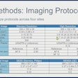
Finally, read more about the inconsistency in the imaging protocols used to assess scaphoid fractures in a trauma setting.


