Gallium-68 (Ga-68) DOTATATE PET could have a new role in helping refine radiation treatment volumes in patients with recurrent pituitary tumors, a team at the University of Wisconsin in Madison has reported.
In a pilot study, Ga-68 DOTATATE PET altered radiation treatment volumes by nearly 20% compared with MRI alone, noted lead author Bradley Eckelmann, MD, PhD, and colleagues.
"Ga-68 DOTATATE PET, when added to MRI for treatment planning, identifies additional target volume and potentially excludes non-avid tissue contoured on MRI," the group wrote. The study was published April 23 in Clinical and Translational Radiation Oncology.
Radiation therapy is recommended by consensus guidelines in patients when pituitary tumors are refractory or not amenable to surgery or medical therapy, the authors explained. MRI is the current standard for radiation planning, yet postsurgical changes can make it challenging to delineate recurrent or residual disease, especially in patients who have undergone multiple prior surgeries, the group added.
Ga-68 DOTATATE PET, which was approved in the U.S. in 2016 for imaging neuroendocrine tumors, targets somatostatin receptors, which are highly expressed in pituitary adenomas. In this study, the researchers hypothesized that adding Ga-68 DOTATATE PET imaging to standard MRI could more accurately identify treatment volumes compared with MRI alone.
The group identified seven patients with recurrent pituitary adenoma who underwent both MRI and Ga-68 DOTATATE PET as part of radiation treatment planning at their institution in 2023 and 2024. All patients had prior surgery -- six subtotal resections and one gross total resection -- and six underwent simultaneous PET/MRI scans. Four radiation oncologists and one resident independently contoured gross tumor volumes on each modality.
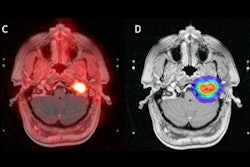
![A 53-year-old patient (patient number four) with a recurrent pituitary adenoma with extension of a cystic component of disease to the medial temporal lobe apparent on MRI (contoured in blue), and extension of disease to the left sphenoid bone and orbital apex apparent on [68Ga]Ga-DOTA-TATE (contoured in yellow).](https://img.auntminnie.com/mindful/smg/workspaces/default/uploads/2026/04/pituitary-tumor.QGsEnyB4bU.jpg?auto=format%2Ccompress&dpr=2&fit=max&q=70&w=700) A 53-year-old patient (patient number four) with a recurrent pituitary adenoma with extension of a cystic component of disease to the medial temporal lobe apparent on MRI (contoured in blue), and extension of disease to the left sphenoid bone and orbital apex apparent on [68Ga]Ga-DOTA-TATE (contoured in yellow).Clinical and Translational Radiation Oncology
A 53-year-old patient (patient number four) with a recurrent pituitary adenoma with extension of a cystic component of disease to the medial temporal lobe apparent on MRI (contoured in blue), and extension of disease to the left sphenoid bone and orbital apex apparent on [68Ga]Ga-DOTA-TATE (contoured in yellow).Clinical and Translational Radiation Oncology
Average PET volumes were both larger and smaller than average MRI volumes for different patients. The range of differences between average MRI and PET volumes was −3.81 cm3 to +4.34 cm3. Lastly, there was a median absolute difference between average PET and average MRI volume of 2.24 cm3, which corresponds to a median relative percent difference of 20.3%, according to the authors.
“Our findings demonstrate that Ga-68 DOTATATE PET in combination with MRI may refine target delineation relative to MRI alone, as it identifies both additional volume and may exclude non-avid regions contoured on MRI,” the group wrote.
The researchers noted limitations, namely the small seven-patient cohort, the retrospective design, and the absence of long-term local control and toxicity data. They also noted that a small proportion of pituitary tumors do not express the specific somatostatin receptor that DOTATATE binds to and that DOTATATE uptake in normal pituitary tissue can complicate image interpretation.
Nonetheless, this is one of the first known comparisons of Ga-68 DOTATATE PET-based and MRI-based radiation treatment volumes for pituitary adenoma patients, the team explained, and the results warrant further investigation.
"Prospective studies with larger patient cohorts are needed to validate our findings and evaluate the impact of PET-based planning on clinical outcomes," Eckelmann and colleagues concluded.
The full study is available here.